

Overview of Macular Corneal Dystrophy
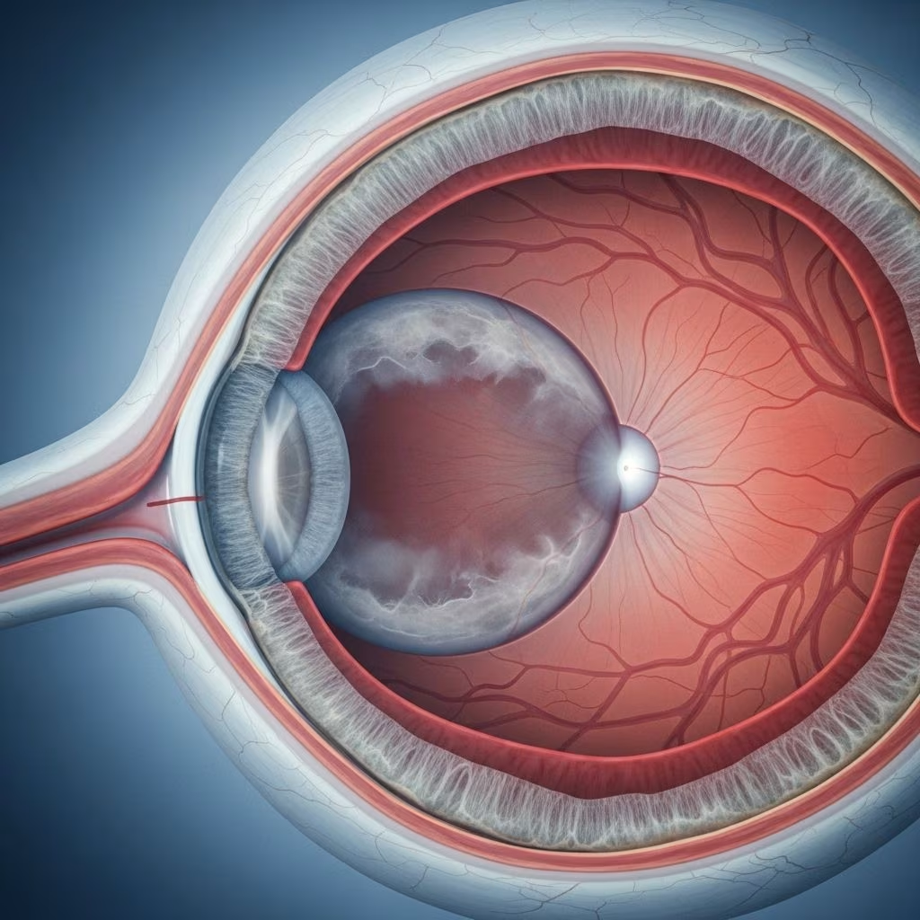
Macular corneal dystrophy is a rare, hereditary eye disease that causes cloudiness and opaque spots in the central part of the cornea. This disease is a type of stromal corneal dystrophy and leads to progressive haziness in vision, often becoming severe over time. The primary underlying cause of macular corneal dystrophy is a mutation in the CHST6 gene, which is responsible for the sulfation of keratan molecules in the cornea. The disease only manifests if the mutated gene is inherited from both parents, making it an autosomal recessive condition.
- Category: Stromal corneal dystrophy
- Inheritance: Autosomal recessive (requires both parents to carry the mutation)
- Prevalence: Fewer than 1 in 10,000 people in the United States
- Key gene: CHST6
Symptoms of Macular Corneal Dystrophy
Symptoms usually begin during the first or second decade of life, though onset may be delayed until as late as the sixth decade. As the disease progresses, vision becomes increasingly impaired due to the expanding opacity and haze in the cornea.
- Decreased visual acuity: Ranging from mild blurring to severe low vision (average visual acuity can range between 20/250 to 20/400)
- Eye irritation and pain: Caused by recurrent erosions and inflammation
- Photophobia: Increased sensitivity to light
- Reduced corneal sensitivity: Due to recurrent erosions
- Progressive corneal haziness: Ill-defined gray-white opacities in the stroma, starting centrally and spreading outward
The corneal opacities merge and expand over time. Anterior stromal deposits can also lead to irregular astigmatism, and continued disease progression results in thinning of the central cornea, as well as possible involvement of the Descemet’s membrane and endothelium.
Visual Progression Table
| Stage | Key Symptoms | Impact on Vision |
|---|---|---|
| Early | Ill-defined gray-white opacities, mild haze | Minimal to moderate visual blurring |
| Mid | Expansion and merging of opacities, possible astigmatism | Increasing visual impairment, photophobia, irritation |
| Late | Severe stromal haze, thinning, Descemet/endothelium involvement | Severe vision loss, possible blindness |
Causes and Genetic Basis
The primary cause of macular corneal dystrophy is a mutation in the CHST6 gene located on chromosome 16. This gene encodes the enzyme carbohydrate (N-acetylglucosamine 6-O) sulfotransferase 6, which catalyzes sulfate transfer to keratan sulfate in the cornea. Mutations result in abnormal accumulation of glycosaminoglycans within the corneal stroma, keratocytes, and sometimes the endothelium, producing the hallmark cloudy opacities and haze.
- Inheritance: Autosomal recessive—must inherit the mutation from both parents
- Immunophenotypes: Variants based on keratan sulfate presence
- Type I: No detectable keratan sulfate in cornea or serum
- Type II: Normal keratan sulfate in cornea and serum
- Type IA: No antigenic keratan sulfate in serum, but present in keratocytes
- Differences between immunophenotypes are not clinically significant
Risk Factors for Macular Corneal Dystrophy
- Family history: Having close relatives with the condition strongly increases risk
- Ethnicity: Certain populations may have higher rates, though the disease is rare globally
- Genetics: Inheritance of two defective copies of the CHST6 gene
There are currently no known environmental risk factors that cause macular corneal dystrophy, as the disease is entirely genetic.
Potential Complications
As macular corneal dystrophy advances, complications may develop—especially if left untreated. Opacities and thinning of the cornea not only diminish vision but can also create vulnerability to other eye problems.
- Severe vision loss or blindness
- Corneal scarring
- Corneal ulcers
- Glaucoma
- Astigmatism (irregular corneal curvature)
- Cataracts
- Endophthalmitis (serious inner ocular infection)
When to Seek Medical Attention
Early detection is key. You should contact an eye care specialist if you experience:
- Progressive blurred vision or visual haze
- Increased light sensitivity
- Unexplained eye pain or irritation
- Reduced ability to distinguish detail or colors
Prompt evaluation helps rule out other causes of corneal opacity and allows timely intervention to preserve vision.
Diagnosis of Macular Corneal Dystrophy
Diagnosis begins with a thorough eye examination. Key diagnostic techniques include:
- Slit lamp examination: Reveals characteristic stromal haze and opacities
- Assessment of corneal curvature, thickness, and astigmatism
- Measurement of visual acuity: Monitoring progression
- Genetic testing: Identifies mutations in CHST6 and confirms diagnosis
- Imaging: Scheimpflug and other imaging may be used to rule out other thinning disorders like keratoconus
A complete ocular history and periodic follow-up are essential for monitoring progression and determining the best timing for intervention. Genetic counseling may be recommended for affected families.
Treatment Options for Macular Corneal Dystrophy
There is no medical cure for the underlying genetic defect in macular corneal dystrophy. However, treatment focuses on:
- Symptom management
- Slowing progression
- Restoring vision through surgical and supportive means
Main Treatments
- Corneal transplantation (keratoplasty): The primary treatment for severe cases; may include partial (lamellar) or full-thickness (penetrating) keratoplasty.
- Eye drops and lubricants: Used to manage irritation and discomfort; cannot cure the condition.
- Protective eyewear: For photophobia and to prevent further trauma.
Regenerative techniques and gene therapies remain experimental but hold future promise.
Outlook and Prognosis
Macular corneal dystrophy tends to be a progressive condition, but effective treatment can restore considerable vision. The prognosis following corneal transplantation is generally favorable, though repeat procedures may be required in some cases due to longer-term recurrence of dystrophy or rejection.
- Vision can often be significantly improved with timely transplantation
- Recurrence is possible: Ongoing follow-up is needed
- Lifelong monitoring is recommended for optimal outcomes
Prevention Strategies
Because macular corneal dystrophy is strictly a genetic disorder, direct prevention is currently impossible. However, certain steps can help mitigate risk and guide affected families:
- Genetic counseling: For families with known history
- Early screening in siblings and offspring
- Prompt evaluation at first symptom onset
Frequently Asked Questions (FAQ)
What is macular corneal dystrophy?
Macular corneal dystrophy is a rare inherited disease that causes clouding and opaque spots in the central cornea, progressively impairing vision.
Is macular corneal dystrophy curable?
No direct medical cure exists. However, corneal transplantation is an effective treatment to restore vision in most cases.
How is macular corneal dystrophy diagnosed?
Diagnosis is through eye examination (slit lamp), assessment of visual acuity, corneal imaging, and confirmation by genetic testing for the CHST6 mutation.
Are there long-term complications?
Untreated, the disease can cause severe vision loss, corneal scarring, ulcers, glaucoma, and other serious eye conditions.
Can macular corneal dystrophy be prevented?
No, it cannot be prevented due to its genetic nature. Family genetic counseling and early screening are recommended for at-risk individuals.
Summary
Macular corneal dystrophy is a rare, autosomal recessive disease that damages the corneal stroma, causing vision-impairing haze and opacities. Symptoms begin early and worsen, with possible complications such as ulcers, glaucoma, and blindness. Diagnosis is through clinical examination and genetic testing for CHST6 mutations. While there is no cure, vision can often be restored with corneal transplantation. Genetic counseling, early detection, and routine monitoring are advised for affected families to maximize visual outcomes and minimize long-term risks.
References
- https://eyewiki.org/Macular_Corneal_Dystrophy
- https://pmc.ncbi.nlm.nih.gov/articles/PMC4194335/
- https://www.healthline.com/health/eye-health/macular-corneal-dystrophy
- https://www.healthline.com/health/eye-health/stages-of-macular-degeneration
- https://www.uc.edu/news/articles/legacy/healthnews/2006/09/uc-health-line–diet-can-help-prevent-age-related-macular-degeneration.html
- https://www.medicalnewstoday.com/articles/macular-degeneration-foods-to-avoid
- https://www.eyesiteonwellness.com/bcbstx/en/
- https://www.britannica.com/science/macular-degeneration
- https://my.clevelandclinic.org/health/diseases/corneal-dystrophy