

Microaneurysms in Diabetic Retinopathy: An In-Depth Guide
Microaneurysms represent one of the earliest clinical signs of diabetic retinopathy, a potentially vision-threatening complication of diabetes. Understanding their role in the progression of retinal disease is crucial for anyone seeking to protect their vision and manage diabetes effectively.
Contents
- About Microaneurysms
- Microaneurysms vs. Hemorrhages
- Impact on Vision
- Causes and Mechanisms
- Stages of Diabetic Retinopathy
- Symptoms and Signs
- Diagnosis and Assessment
- Treatment and Management
- Prevention Strategies
- Frequently Asked Questions
What Are Microaneurysms from Diabetic Retinopathy?
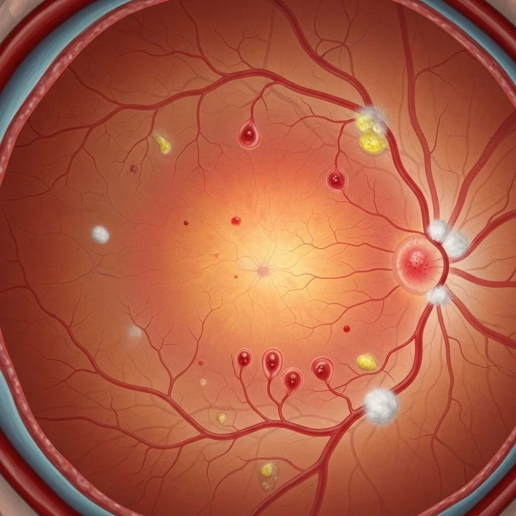
Microaneurysms are tiny areas where the blood vessels in the retina swell and bulge, often appearing as minute red dots during retinal examination. These represent focal weaknesses in the vessel walls caused by high blood sugar related damage and are the first visible signs of diabetic retinopathy in most cases.
As diabetes progresses, these microaneurysms can leak fluid or blood, indicating increasing damage to retinal vasculature. The detection of microaneurysms is a key marker for clinicians and sets the stage for evaluating the risk and management of diabetic eye disease.
Microaneurysms vs. Hemorrhages
While both microaneurysms and hemorrhages involve the breakdown of small retinal blood vessels, they differ significantly:
- Microaneurysms: Small, bulging areas caused by focal weakening of vessel walls; may leak fluid or blood but remain generally dot-like.
- Retinal Hemorrhages: Larger areas of bleeding into the retina. “Dot and blot” hemorrhages result from microaneurysms rupturing and blood pooling within the retina’s nerve fiber layer, sometimes appearing flat and feathery (blot hemorrhages).
These events increase in frequency and severity as diabetic retinopathy progresses.
What Will Happen to Your Vision If You Have a Microaneurysm?
Most microaneurysms do not immediately affect vision, especially if they are located outside the macula—the central part of the retina responsible for sharp, detailed vision.
However, when microaneurysms leak into the macula, fluid and proteins can cause macular edema. The macula contains the greatest density of photoreceptors responsible for central vision and focus. Swelling here can impede sharp vision, sometimes leading to blurry vision or vision loss. About 1 in 15 people with diabetes will develop diabetic macular edema.
- If microaneurysms leak elsewhere in the retina (outside the macula), you typically have no symptoms, but their presence signals increasing risk for vision complications.
- Untreated damage progressing from microaneurysms can eventually affect larger areas of the retina, leading to vision problems and even blindness in severe cases.
Causes and Mechanisms of Microaneurysm Formation
Microaneurysm development results primarily from chronic high blood sugar (hyperglycemia) in diabetes. Over time, elevated glucose levels damage the cells lining small blood vessels throughout the body, especially in the retina.
Key mechanisms include:
- Blood Vessel Wall Damage: High glucose levels compromise the integrity of capillary endothelial cells, causing them to swell and weaken.
- Increased Vascular Permeability: Vessel walls become leaky, resulting in fluid and protein escaping into retinal tissue.
- Local Inflammation: Diabetic retinopathy promotes inflammation, accelerating vessel wall damage and microaneurysm formation.
Microaneurysm appearance marks the first stage of diabetic retinopathy, often preceding permanent changes to vision.
Stages of Diabetic Retinopathy
Diabetic retinopathy typically progresses through four main stages, each reflecting escalating damage to blood vessels and retinal tissue.
| Stage | Description | Key Findings |
|---|---|---|
| Mild Nonproliferative Retinopathy | First stage; presence of microaneurysms. | Microaneurysms, occasional dot hemorrhages. |
| Moderate Nonproliferative Retinopathy | Vessel blockages begin; more hemorrhages. | Blocked vessels, development of “cotton-wool spots,” hard exudates. |
| Severe Nonproliferative Retinopathy | Widespread narrowing of retinal vessels, lowered oxygen supply. | Extensive hemorrhages, subtle vascular changes. |
| Proliferative Retinopathy | Advanced stage; new abnormal vessels grow. | Neovascularization, severe hemorrhage, retinal detachment risk. |
Symptoms and Signs of Diabetic Retinopathy (Including Microaneurysms)
Microaneurysms themselves rarely cause symptoms unless leakage leads to macular edema. As retinopathy worsens, symptoms can range from mild to severe:
- Early Stages: Usually asymptomatic; microaneurysms detected only during eye exams.
- Intermediate Stages: Blurred vision (especially if or when macular edema develops), floating spots, or mild distortion.
- Advanced Stages: Sudden, painless vision loss; floaters (black spots), flashing lights, or dark areas in visual field; result from hemorrhage or retinal detachment.
Specific signs detected on eye examination include:
- Capillary microaneurysms (first sign)
- Dot and blot retinal hemorrhages
- Hard exudates: yellow retinal particles suggest chronic edema
- Cotton-wool spots: white, fuzzy-edged areas indicating microinfarction
- Macular edema: retinal swelling and blurred retinal layers
- Venous dilation and intraretinal microvascular abnormalities
In advanced “proliferative” stages, abnormal new vessels may grow and leak, causing further damage and greater risk of permanent vision loss.
Diagnosis and Assessment of Microaneurysms
- Regular Eye Exams: Microaneurysms are identified using specialized tests such as fundus photography, fluorescein angiography, and optical coherence tomography (OCT).
- Clinical Grading: Ophthalmologists use the number and location of microaneurysms to stage diabetic retinopathy and assess progression risk.
- Macular Edema Detection: Advanced imaging identifies macular thickening from fluid leakage, allowing for early intervention.
Early detection is crucial because symptoms often appear only after substantial damage has occurred.
Treatment and Management of Microaneurysms and Retinopathy
Treatment is tailored to the stage and severity of retinopathy. Microaneurysms indicate the need to intensify diabetes care and monitor closely for progression.
Primary goals: slow disease, reduce fluid leakage, and protect vision.
- Tight Blood Sugar Control: Maintaining near-normal glucose levels can slow retinal vessel damage and the appearance of new microaneurysms.
- Blood Pressure Management: High blood pressure amplifies vessel damage; treatment is critical.
- Regular Ophthalmologic Monitoring: Frequent eye exams allow early detection of changes and timely treatment adjustments.
- Medical Therapies:
- Anti-VEGF injections for macular edema
- Corticosteroid injections where chronic inflammation is present
- Laser photocoagulation to seal leaking microaneurysms and prevent progression
- Surgical Intervention: Advanced cases with retinal detachment or severe bleeding may require vitrectomy or other procedures.
Prompt treatment of microaneurysm-related leakage in the macula (macular edema) can help prevent lasting central vision loss.
Prevention Strategies
- Routine Diabetes Management: Keep blood sugar, blood pressure, and cholesterol under control.
- Annual Diabetic Eye Exams: Early detection of microaneurysms and other changes enables rapid intervention.
- Healthy Lifestyle: Diet, exercise, weight management, and smoking cessation all reduce vascular risks.
- Medication Compliance: Take diabetes medications consistently as prescribed.
The risk of vision-threatening complications is markedly reduced with early diagnosis and prompt, sustained management.
Frequently Asked Questions (FAQs)
Q: Are microaneurysms in the eye reversible?
A: Microaneurysms themselves usually do not reverse once formed, but controlling underlying diabetes and treating retinal leakage can prevent further damage.
Q: Will I lose my vision if I have microaneurysms?
A: Most people do not experience symptoms from microaneurysms alone, unless leakage leads to macular edema. Prompt diabetes and eye care can greatly reduce the risk of vision loss.
Q: How often should I have my eyes checked if I have diabetes?
A: Eye experts recommend annual dilated eye exams for all people with diabetes unless otherwise advised, as early changes can only be detected with clinical tests.
Q: What is the difference between microaneurysms and retinal hemorrhages?
A: Microaneurysms are tiny bulges in vessel walls, often seen as red dots; retinal hemorrhages are actual leaks or pools of blood in retinal layers resulting from ruptured vessels, and are typically larger.
Q: Are microaneurysms only found in diabetic retinopathy?
A: While most commonly associated with diabetic retinopathy, microaneurysms may also occur in other conditions that damage retinal vessels, though they are rare without systemic disease.
Summary Table: Key Facts About Diabetic Retinopathy Microaneurysms
| Feature | Description |
|---|---|
| Definition | Tiny bulges or weak spots in retinal blood vessel walls; first sign of diabetic retinopathy. |
| Symptoms | Most are asymptomatic; may cause vision changes if leading to macular edema. |
| Diagnosis | Detected in retinal imaging during comprehensive eye exams. |
| Treatment | Control of blood sugar, blood pressure, specific retinal treatments (injections, laser). |
| Risk Factors | Poorly controlled diabetes, high blood pressure, high cholesterol, long duration of diabetes. |
Conclusion
Microaneurysms are a critical early marker of diabetic retinopathy—a progressive eye disease linked with diabetes. Though they seldom produce symptoms on their own, their detection signals the need for close monitoring and proactive diabetes management to preserve vision. Timely intervention and adherence to medical guidance can significantly reduce the risk of vision loss related to diabetic retinopathy.
References
- https://www.healthline.com/health/diabetes/diabetic-retinopathy-microaneurysms
- https://www.mayoclinic.org/diseases-conditions/diabetic-retinopathy/symptoms-causes/syc-20371611
- https://www.merckmanuals.com/professional/eye-disorders/retinal-disorders/diabetic-retinopathy
- https://my.clevelandclinic.org/health/diseases/8591-diabetic-retinopathy
- https://www.uchicagomedicine.org/conditions-services/ophthalmology/diabetic-retinopathy
- https://www.orbis.org/en/avoidable-blindness/diabetic-retinopathy
- https://www.ncbi.nlm.nih.gov/books/NBK560805/
- https://www.rvscny.com/patient-eduction/conditions-we-treat/diabetic-retinopathy/