

Thrombotic Microangiopathy: Causes, Impact on Kidneys, and Management
Thrombotic Microangiopathy (TMA) is a rare, serious condition defined by injury to the smallest blood vessels—often affecting organs like the kidney and the brain. This comprehensive article explores TMA from its fundamental pathology and causes to diagnostic methods, treatments, and prognosis, with a focus on kidney health.
The content is organized to guide readers through understanding the disease, identifying its core features and subtypes, and learning about modern management approaches.
What Is Thrombotic Microangiopathy?
TMA stands for thrombotic microangiopathy—a term that translates to a small blood vessel problem involving abnormal clot formation. In TMA, blood clots form in tiny vessels throughout the body, most notably in the kidneys, causing organ damage and potentially life-threatening complications.
Key features of TMA include:
- Microangiopathic hemolytic anemia: Red blood cells are damaged as they pass through small, blocked vessels.
- Thrombocytopenia: Platelet counts drop as platelets are consumed forming clots.
- Organ injury: Especially acute kidney injury (AKI), but can also affect the brain and other vital organs.
How Does TMA Affect Kidney Function?
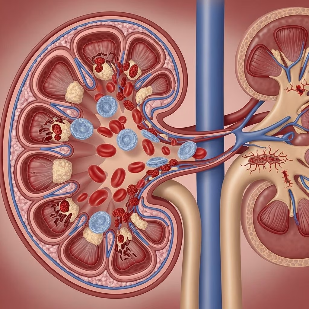
In a healthy kidney, tiny vessels called capillaries are lined by a smooth layer of endothelial cells, which facilitate blood flow, filtration, and waste removal. Damage to these cells in TMA slows blood movement, distorts vessel walls, and leads to a cascade where red blood cells rupture and platelets aggregate into clots.
Consequences for the kidney include:
- Blood vessel blockage that impairs oxygen delivery and normal filtering function.
- Decreased red blood cells (anemia) and platelets, which hinders blood clotting and oxygen supply.
- Tissue injury or death in affected kidney segments due to prolonged lack of blood flow.
Microscopic Findings in TMA
Laboratory and microscopic analysis reveals telltale changes:
- Kidney blood vessels may appear filled with pinkish clots under the microscope.
- Areas devoid of normal blood flow become pale and damaged over time.
- Blood samples show deformed red blood cells called schistocytes and reduced platelets.
These findings help differentiate TMA from other causes of kidney dysfunction.
What Causes Thrombotic Microangiopathy?
TMA is not a single disease, but a pattern of injury driven by various underlying disorders. The most common causes can be classified as follows:
| Condition | Abbreviation | Key Features |
|---|---|---|
| Thrombotic Thrombocytopenic Purpura | TTP |
|
| Hemolytic Uremic Syndrome | HUS, Typical & Atypical |
|
| Malignant Hypertension | — |
|
| Disseminated Intravascular Coagulation | DIC |
|
| HELLP Syndrome | HELLP |
|
| Scleroderma Renal Crisis | SRC |
|
Overview of Main TMA Subtypes
Thrombotic Thrombocytopenic Purpura (TTP)
TTP is characterized by severe deficiency of ADAMTS13, the enzyme responsible for cleaving von Willebrand factor. In its absence, large von Willebrand factor multimers promote platelet aggregation, causing microscopic clots.
There are two main forms:
- Acquired TTP: Autoimmune—antibodies block ADAMTS13 function.
- Congenital TTP: Genetic deficiency, often presenting in childhood but can be triggered later (e.g., pregnancy).
Core symptoms include:
- Fatigue due to anemia
- Bruising or petechiae from low platelets
- Neurological symptoms (confusion, headache)
- Acute kidney injury
Hemolytic Uremic Syndrome (HUS)
HUS is most commonly triggered by infection with Shiga toxin–producing E. coli, leading to bloody diarrhea followed by kidney injury.
Subtypes:
- Typical HUS: Detectable Shiga toxin or pathogenic E. coli in stool.
- Atypical HUS: Caused by alternative complement pathway dysregulation (often genetic or autoimmune).
Symptoms include:
- Acute kidney injury/failure
- Hemolytic anemia
- Low platelet count
- Gastrointestinal symptoms (mostly in typical HUS)
Other Causes: Atypical Presentations and Systemic Conditions
Beyond TTP and HUS, TMA can result from:
- Malignant hypertension: Extremely high blood pressure causing vascular injury.
- DIC: Widespread clot formation due to sepsis, trauma, or malignancy.
- HELLP syndrome: Pregnancy complication with hemolysis and liver involvement.
- Scleroderma renal crisis: Seen in systemic scleroderma.
For each of these, clinical suspicion and laboratory findings are key for diagnosis and treatment planning.
Risk Factors and Susceptible Populations
Anyone can develop TMA, but higher risk exists for certain groups:
- Children—especially under age 5 (more at risk for HUS following bacterial infection)
- Adults—more likely to develop TTP and atypical HUS
- Pregnant women—at risk for HELLP syndrome and TTP
- Individuals with autoimmune diseases or inherited genetic mutations
Signs, Symptoms, and Clinical Presentation
Symptoms vary by underlying cause and severity, but most patients present with:
- Fatigue and pallor (related to anemia)
- Easy bruising, petechiae, and nosebleeds (due to thrombocytopenia)
- Acute kidney injury: swelling, decreased urine output, elevated creatinine
- Confusion, headaches, or seizures (when the brain is affected)
- Abdominal pain or diarrhea (especially in HUS)
Diagnostic Approach to TMA
TMA diagnosis is multifaceted:
- Lab Tests:
- Complete blood count (showing anemia, low platelets)
- Peripheral blood smear (schistocytes are a hallmark)
- Renal function tests (creatinine, BUN, electrolytes)
- ADAMTS13 levels/activity
- Complement system analysis for atypical HUS
- Stool tests for Shiga toxin in typical HUS
- Imaging: Ultrasound and CT scans may assess kidney damage or complications.
- Renal biopsy: Reveals classic microangiopathic
Modern Treatment Strategies for TMA
The treatment of TMA depends on its subtype and underlying cause:
- TTP:
- Urgent plasma exchange to remove autoantibodies and replace ADAMTS13.
- Corticosteroids and sometimes immunosuppressive agents.
- Typical HUS:
- Mainly supportive care (fluids, dialysis if necessary).
- Antibiotics usually avoided to minimize toxin release.
- Atypical HUS:
- Eculizumab (monoclonal antibody that inhibits complement system, highly effective for complement-driven HUS).
- Other causes:
- Treating the underlying disorder (e.g., controlling hypertension, managing DIC).
Early diagnosis and rapid intervention are critical to prevent organ failure and long-term complications.
Prognosis and Long-Term Outcomes
Outcomes depend on the subtype, severity, and speed of treatment initiation:
- Some patients recover fully with rapid, appropriate therapy.
- Others may experience chronic kidney damage, need for ongoing dialysis, or neurological complications.
- Mortality is highest in untreated or severe cases, especially those with underlying systemic disease.
Recognizing TMA early and understanding its multi-system impact is crucial for improving prognosis.
Frequently Asked Questions About Thrombotic Microangiopathy
Q: Can TMA happen to anyone?
A: Yes, but certain groups—children, adults with underlying conditions, pregnant women—have higher risks, depending on the TMA subtype.
Q: What are the hallmark symptoms of TMA?
A: Fatigue, bruising, decreased kidney function, abnormal blood tests (anemia, low platelets), and sometimes neurological symptoms like confusion or seizures.
Q: How is TMA diagnosed?
A: Diagnosis combines clinical suspicion with lab tests (CBC, blood smear, kidney function) and sometimes specialized tests for ADAMTS13 activity or Shiga toxin detection.
Q: Is TMA treatable?
A: Many cases are treatable or manageable, especially if recognized early. Treatment varies by cause and can include plasma exchange, complement inhibitors, supportive care, and addressing underlying systemic conditions.
Q: What is the outlook for people with TMA?
A: Prognosis varies. Some recover completely; others develop chronic kidney damage or experience life-threatening complications if treatment is delayed. Early intervention is essential.
Q: Can TMA recur?
A: Yes, especially with congenital or genetic forms, or if underlying triggers (autoimmune disease, complement pathway mutations) persist.
Q: Are children more affected by HUS, and adults by TTP?
A: Generally, yes. Preschool-age children are susceptible to typical HUS after bacterial infections, while TTP and atypical HUS are more common in adults.
Q: Is organ failure reversible in TMA?
A: Sometimes. With prompt diagnosis and appropriate therapy, many patients regain kidney function, but some may need long-term dialysis or experience lasting damage.
Key Points to Remember
- TMA is not one disease but a pattern caused by various disorders leading to clot formation in small vessels, especially affecting the kidney.
- Diagnosis requires awareness, specialized testing, and swift action to distinguish underlying causes and initiate treatment.
- Most patients benefit from targeted therapy based on the specific subtype, with supportive care playing a vital role).
- Ongoing research is improving outcomes by expanding understanding of genetic and immune system factors in TMA.
References and Further Reading
- UNC Kidney Center: Thrombotic Microangiopathy (TMA)
- Clinical Journal of the American Society of Nephrology: Thrombotic Microangiopathy and the Kidney
- NIH PubMed: Thrombotic Microangiopathies: A General Approach to Diagnosis and Management
References
- https://unckidneycenter.org/kidneyhealthlibrary/glomerular-disease/thrombotic-microangiopathy-tma/
- https://pubmed.ncbi.nlm.nih.gov/29042465/
- https://pmc.ncbi.nlm.nih.gov/articles/PMC5266569/
- https://www.healthline.com/health/multiple-sclerosis/uti-kidney-problems
- https://onlinelibrary.wiley.com/doi/full/10.1002/ajh.24339
- https://www.upmc.com/services/kidney-disease/conditions/kidney-failure