

Chronic lymphocytic leukemia (CLL) is a form of blood cancer that primarily affects a type of white blood cell known as lymphocytes. Diagnosing and managing CLL requires careful analysis of blood and bone marrow samples. One of the most advanced tools for diagnosing CLL and determining its nature is flow cytometry. This test helps doctors confirm the presence of CLL, characterize its aggressiveness, and make informed decisions about treatment. This in-depth article will explain what CLL flow cytometry entails, how the test works, what the results mean, and its role in the overall care of people with CLL.
What Is Flow Cytometry?
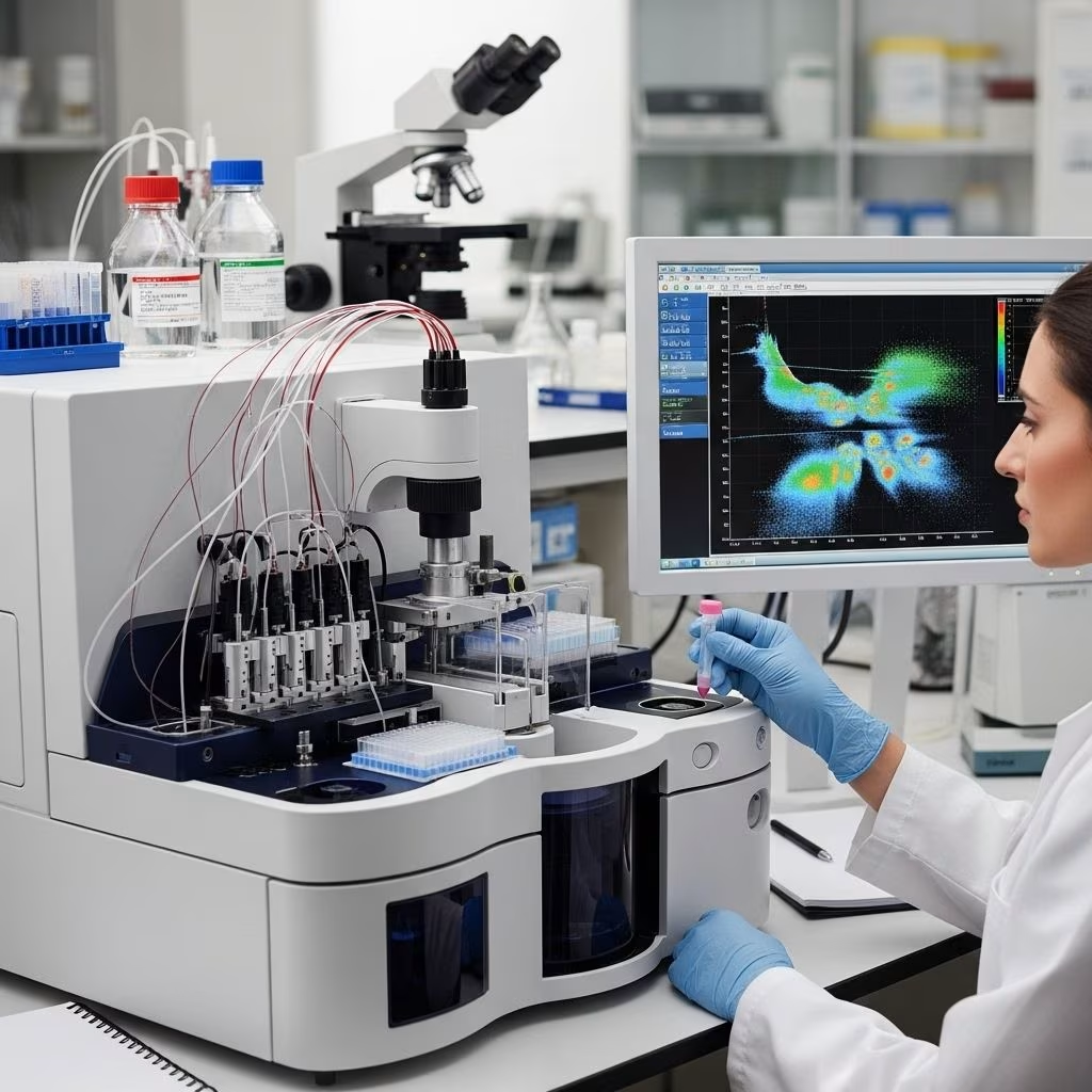
Flow cytometry is a specialized laboratory technique used to measure and analyze the physical and chemical characteristics of cells or particles in a fluid as they pass through at least one laser beam. The flow cytometer—the instrument used for this test—generates data about individual cells, enabling doctors to identify abnormal cells and marker patterns that are indicators of diseases like leukemia.
- Applications: Primarily used to examine abnormal populations of cells, including cancer cells, immune cells, and sometimes even sperm cells.
- Sample Types: Typically performed on blood and bone marrow samples, but it can be adapted for use on other bodily fluids.
Flow cytometry allows for:
- Analyzing numerous characteristics at the single-cell level
- Rapidly processing thousands of cells per second
- Identifying specific cell types by detecting unique markers on cell surfaces
- Determining the lineage of abnormal cells, such as distinguishing B-cells in CLL from other cell types in leukemias or lymphomas
How Does Flow Cytometry Work?
The flow cytometry process involves several crucial steps:
- Sample Collection: Blood or bone marrow is drawn from the patient.
- Lab Preparation: The sample is treated with special antibodies tagged with fluorescent dyes (fluorochromes) that bind to specific cell markers.
- Creating a Cell Suspension: The cells are suspended in a fluid and loaded into the flow cytometry machine.
- Laser Detection: Cells pass one by one through a laser beam. The fluorescence emitted by bound antibodies and the way light scatters as it passes through the cells provide information about cell size, complexity, and the presence of specific proteins or antigens.
- Data Analysis: Detectors capture light signals, converting them into digital data displayed as graphs (dot plots or histograms) for expert interpretation.
Experts can analyze the pattern of markers and other cell properties to distinguish between normal and abnormal cell populations—a crucial step in diagnosing CLL and differentiating it from other blood disorders.
How Is Flow Cytometry Used in CLL Diagnosis?
Flow cytometry is considered a cornerstone in diagnosing chronic lymphocytic leukemia. Here is why this test is so critical in CLL care:
- Confirmation of CLL: CLL is characterized by an increased number of certain white blood cells (B-cell lymphocytes) that display a specific profile of surface markers (such as CD19, CD20, CD5, and CD23). Flow cytometry can verify this marker pattern, which distinguishes CLL from other similar blood cancers.
- Differential Diagnosis: Morphological analysis alone (looking at cells under a microscope) cannot always differentiate CLL from other diseases with similar appearances, like mantle cell lymphoma. Flow cytometry aids in making a definitive diagnosis by identifying the exact set of markers.
- Assessing Aggressiveness: The presence or absence of certain markers can signal how aggressive the CLL may be. Some markers are linked to faster progression and can guide treatment decisions.
- Staging Disease: Quantifying abnormal lymphocytes and characterizing their markers helps determine the disease stage, which is vital for planning therapy and estimating prognosis.
- Monitoring Progression: Flow cytometry can be repeated over time to monitor changes in the number and characteristics of lymphocytes, providing insights into disease progression or response to therapy.
Markers Evaluated in CLL Flow Cytometry
| Marker | Also Known As | Relevance in CLL |
|---|---|---|
| CD19 | B-cell marker | Indicative of B lymphocytes; commonly found in CLL cells. |
| CD5 | T-cell marker also seen on CLL B-cells | Pivotal in CLL diagnosis due to its co-expression with B-cell markers. |
| CD23 | B-cell activation marker | Helps distinguish CLL from mantle cell lymphoma, where CD23 is usually absent. |
| CD20 | B-cell marker | Typically seen in lower density in CLL than other B-cell lymphomas. |
| Light Chains (kappa/lambda) | Immunoglobulin chains | Indicates clonality of the lymphocyte population, helping to confirm malignancy. |
This detailed profile assists in producing a comprehensive diagnostic picture for each patient with suspected CLL.
What Information Does Flow Cytometry Provide?
Flow cytometry provides a wealth of information to oncologists and hematologists:
- Cell Characteristics: Size, internal complexity, and surface receptor composition.
- DNA Content: Measurement of DNA can highlight abnormal patterns seen in malignant cells.
- Protein Expression: Certain proteins are only found on CLL cells, and their detection helps to confirm diagnosis.
- Clonal Identification: CLL cells usually originate from a single clone (genetically identical cell group), which flow cytometry can identify by showing restriction to either kappa or lambda light chains.
- Cellular Subset Analysis: Flow cytometry can separate lymphocyte subsets (B-cells vs T-cells) and further subtype them based on additional markers.
The test also helps determine the percentage of abnormal lymphocytes compared to the healthy population, which is relevant for staging and prognosis.
Other Diagnostic Tests Used in CLL
Although flow cytometry is a critical part of the diagnostic process for CLL, doctors may order additional tests to get a complete picture of the disease:
- Complete Blood Count (CBC): Measures the number and types of blood cells.
- Peripheral Blood Smear: Allows pathologists to examine the shape and appearance of blood cells under a microscope.
- Bone Marrow Biopsy: Sometimes required to confirm diagnosis or assess bone marrow involvement.
- Genetic Testing (FISH): Detects chromosomal abnormalities associated with CLL and helps predict treatment response.
- Immunophenotyping: A lab technique closely related to flow cytometry used to study protein expressions on cell surfaces.
Doctors integrate the results of all these tests to confirm CLL, rule out other conditions, and determine the best treatment strategy.
What Happens After the CLL Flow Cytometry Test?
The results of flow cytometry are interpreted in the context of all other laboratory and clinical findings. Key post-test considerations include:
- Confirming the CLL diagnosis and distinguishing it from other forms of leukemia and lymphoma
- Determining CLL stage based on the amount and distribution of the abnormal lymphocytes
- Guiding treatment decisions, such as the urgency and type of therapy needed
- Assessing prognosis by identifying features associated with faster or slower disease progression
Once the diagnosis and staging are established, your oncology team can discuss therapeutic options, monitoring schedules, and supportive care based on your unique disease profile.
Frequently Asked Questions (FAQs)
What is the purpose of CLL flow cytometry?
CLL flow cytometry is used to detect and measure specific markers on lymphocytes, confirming a diagnosis of chronic lymphocytic leukemia, providing staging information, and helping choose treatment.
How is flow cytometry different from other blood tests?
Unlike general blood tests or blood smears, flow cytometry analyzes hundreds to thousands of cells individually to determine cell lineage and marker patterns. It provides detailed information about the cell’s protein expression and DNA, which cannot be determined by routine tests.
How long does the test take, and when can I expect results?
The flow cytometry procedure is quick, often completed within a few hours at the laboratory, but sample transport and analysis may take a couple of days. Results are typically available within a week.
Why do I need genetic testing like FISH along with flow cytometry?
While flow cytometry diagnoses and characterizes CLL, genetic tests such as FISH (fluorescence in situ hybridization) detect chromosomal abnormalities that further refine prognosis and treatment recommendations.
Will I need repeat flow cytometry during my CLL journey?
Repeat flow cytometry is sometimes needed after treatment, during disease monitoring, or when there is suspicion of disease progression or transformation.
What do the terms CD19, CD5, and CD23 mean?
These are protein markers expressed on the surface of lymphocytes. The co-expression of CD19 and CD5 is characteristic of CLL, while CD23 helps differentiate CLL from similar diseases.
Key Takeaways
- Flow cytometry is vital for diagnosing and staging CLL. It enables precise identification of cancerous lymphocytes based on unique surface markers.
- The test informs treatment and prognosis. The marker profile can predict disease aggressiveness, shaping therapy choices and follow-up plans.
- Flow cytometry is often combined with other tests. Comprehensive CLL diagnosis and management require integrating results from CBC, bone marrow biopsies, and genetic tests.
- Repeat testing may be required during treatment and to monitor for disease changes.
Additional Resources
- Leukemia & Lymphoma Society: Information on CLL and latest research.
- American Cancer Society: Guidance on diagnosis and treatment of CLL.
- Cancer Research UK: Understanding genetic and lab tests in CLL.
References
- https://www.patientpower.info/chronic-lymphocytic-leukemia/cll-flow-cytometry-test
- https://www.healthline.com/health/leukemia/cll-flow-cytometry
- https://www.medicalnewstoday.com/articles/cll-flow-cytometry
- https://www.healthline.com/health/cll/cll-diagnosis-criteria
- https://pmc.ncbi.nlm.nih.gov/articles/PMC5817234/
- https://www.medicalnewstoday.com/articles/cll-diagnosis
- https://www.ncbi.nlm.nih.gov/books/NBK586209/
- https://www.nature.com/articles/leu2015313