

\n\n
Gum cancer is a rare but serious type of oral cancer that arises from the tissues of the gums. Early signs are often mistaken for other common gum diseases, making timely diagnosis and treatment crucial for recovery. Understanding the risk factors, warning signs, diagnostic procedures, and treatment options can help improve outcomes and patient well-being.
\n\n
What Is Gum Cancer?
\n
Gum cancer, medically known as gingival squamous cell carcinoma, is a malignant tumor that develops from the cells lining the gums. It is classified under the broader group of oral cancers, which represent about 3% of new cancer diagnoses annually. Most cases are squamous cell carcinomas, known for their aggressive nature and tendency to spread quickly.
\n\n
While gum cancer can occur in either the upper or lower gums, the approaches to treatment may differ based on the location and extent of the disease. Early detection is vital, as early-stage gum cancer is highly curable and treatments aim to preserve oral function and appearance as much as possible.
\n\n
Risk Factors for Gum Cancer
\n
Several factors can increase an individual’s risk for developing gum cancer. Understanding these can help with prevention and early intervention.
\n
- \n
- Tobacco Use: Smoking cigarettes, cigars, or pipes, and especially chewing tobacco increases risk significantly.
- Alcohol Consumption: Heavy, regular alcohol intake is another major risk factor.
- Human Papillomavirus (HPV): Certain strains of HPV are associated with a higher risk for oral cancers, including those of the gums.
- Chronic Gum Disease: Ongoing inflammation such as gingivitis or periodontitis can predispose to malignancy.
- Sun Exposure: Chronic sun exposure increases the risk for cancers of the lips and may play a minor role for gums.
- Poor Oral Hygiene: Consistently poor dental care contributes to risk by fostering chronic irritation and infection.
- Age and Sex: Gum cancer is more common in people over 40 years old and slightly more common in men.
- Dietary Factors: Low intake of fruits and vegetables may increase the risk.
\n
\n
\n
\n
\n
\n
\n
\n
\n\n
Symptoms of Gum Cancer
\n
The symptoms of gum cancer can closely resemble those of non-cancerous gum diseases, leading to potential delays in diagnosis. Being alert to signs that do not improve can prompt early evaluation and better outcomes.
\n
- \n
- Persistent gum bleeding without a clear cause or that does not heal
- Pain or discomfort in the gums, especially while chewing or brushing
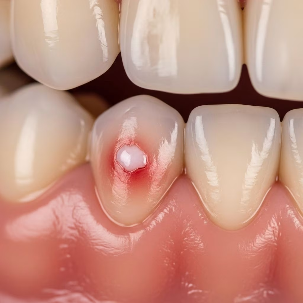
- White, red, or dark patches on the gum tissue
- Unexplained mouth sores or ulcers that do not heal within two weeks
- Gum swelling causing poorly fitting dentures or teeth becoming loose
- Unusual lumps or thickened areas on the gum line
- Cracking, roughness, or crusty areas of the gums
- Ear pain or jaw discomfort not explained by other conditions
\n
\n
\n
\n
\n
\n
\n
\n
\n
Note: Many of these symptoms overlap with other dental problems. Consult a dentist or healthcare provider if they persist for more than two weeks.
\n\n
Causes and Development
\n
Gum cancer begins when genetic mutations in the cells lining the gums cause them to grow uncontrollably, forming tumors or lesions. The majority are squamous cell carcinomas, while a rarer, slower-growing form called verrucous carcinoma also exists. Gum cancer can begin as pre-cancerous changes, such as leukoplakia (gray or white patches) or erythroplakia (red patches), some of which may progress to malignancy over time.
\n\n
How Gum Cancer Spreads
\n
Initially, the cancer may be limited to the outermost layers of gum tissue (carcinoma in situ). Over time, it can invade deeper tissues such as bone and may metastasize (spread) to lymph nodes or other organs if not treated early.
\n\n
How Is Gum Cancer Diagnosed?
\n
Early diagnosis is essential for improving survival and quality of life. Dentists often detect suspicious gum changes during routine checkups, but confirmation requires a detailed evaluation:
\n
- \n
- Physical Examination: The dentist or surgeon examines the gums and oral cavity for abnormal lesions, swelling, or discoloration.
- Biopsy: Removal of a small tissue sample from the affected gums for laboratory analysis to confirm cancer type and stage.
- Imaging Tests: X-rays, CT scans, MRI, or PET scans help determine the size of the tumor, its spread to adjacent structures, and any lymph node involvement.
- Staging: Cancer staging evaluates how deeply the cancer has invaded tissues, whether it has reached the jawbone, and if it has spread to lymph nodes or distant sites. This staging guides treatment planning.
\n
\n
\n
\n
\n\n
| Stage | Description |
|---|---|
| Stage 0 (Carcinoma in situ) | Cancer is limited to the outer layer of gum tissue (epithelium). |
| Stage I | Tumor is less than 2 cm and has not spread to lymph nodes. |
| Stage II | Tumor more than 2 cm but less than 4 cm, no lymph node involvement. |
| Stage III | Tumor larger than 4 cm, or any size with a single lymph node involvement. |
| Stage IV | Cancer has spread to multiple lymph nodes or distant organs. |
\n
\n\n
Treatment Options for Gum Cancer
\n
Treatment for gum cancer depends on the location, size, stage, and general health of the patient. Treatments aim to remove the cancer, minimize the risk of recurrence, and preserve oral function and appearance as much as possible. A multidisciplinary team, including surgeons, oncologists, dentists, reconstructive surgeons, and other specialists, collaborates on individualized treatment plans.
\n\n
Surgical Treatments
\n
- \n
- Wide Local Excision: Removal of the tumor with surrounding healthy tissue to ensure complete cancer excision.
- Mandibulectomy or Maxillectomy: If cancer has invaded the jawbone (mandible) or upper jaw (maxilla), part of the affected bone may be surgically removed.
- Neck Dissection: When there is spread or suspicion of spread to lymph nodes, a neck dissection removes those nodes to prevent further metastasis.
- Reconstructive Surgery: After tumor and bone removal, reconstructive surgery may restore appearance and function, using tissue or bone grafts from other parts of the body and sometimes dental prosthetics.
\n
\n
\n
\n
\n\n
Non-Surgical Treatments
\n
- \n
- Radiation Therapy: High-energy beams are used to destroy cancer cells. It can be an alternative to surgery for small tumors, or used after surgery to reduce recurrence risk.
- Chemotherapy: Powerful medications are used to kill cancer cells, often in combination with radiation for advanced cases, or when surgery is not feasible.
- Targeted Therapy/Immunotherapy: These advanced treatments may be considered for recurrent or metastatic gum cancer, targeting specific cancer cell mechanisms or boosting the immune response.
\n
\n
\n
\n\n
Treatment Goals
\n
- \n
- Remove the tumor completely
- Preserve oral function and appearance
- Prevent recurrence and further spread
\n
\n
\n
\n\n
Special Considerations for Upper vs. Lower Gum Cancers
\n
- \n
- Upper gum cancer may require maxillectomy and reconstruction of the roof of the mouth.
- Lower gum cancer often needs mandibulectomy (removal of part of the jawbone) and possibly neck dissection if lymph nodes are involved.
\n
\n
\n\n
Supportive Care During and After Treatment
\n
- \n
- Speech and Swallowing Therapy: Rehabilitation may be needed after extensive surgery.
- Nutritional Support: Dietitians help maintain nutrition through recovery.
- Dental and Oral Care: Ensuring dental health before and after cancer therapy can reduce complications.
- Psychosocial Support: Counseling can help patients cope with the emotional effects of cancer treatment.
\n
\n
\n
\n
\n\n
Prognosis and Outlook
\n
When detected at an early stage, gum cancers have a high curability rate and good long-term outcomes. Prognosis depends on several factors:
\n
- \n
- Stage at Diagnosis: Earlier stages have better outcomes.
- Tumor Size and Location: Smaller tumors and those limited to the soft tissue are easier to treat.
- Lymph Node Involvement: Spread to lymph nodes lowers the chances of cure but can be managed with aggressive treatment.
- Overall Health: General health and ability to tolerate therapy affect recovery.
\n
\n
\n
\n
\n
Regular dental checkups, early detection, and lifestyle changes such as quitting tobacco and limiting alcohol play an important role in improving survival and preventing gum cancer recurrence.
\n\n
Preventing Gum Cancer
\n
While not all risk factors are avoidable, there are several key measures to reduce the risk of gum cancer:
\n
- \n
- Avoid tobacco and limit alcohol use.
- Maintain proper oral hygiene with regular brushing, flossing, and dental checkups.
- Protect lips and mouth from excessive sun exposure where relevant.
- Eat a healthy diet rich in fruits and vegetables.
- Address dental problems promptly, including ill-fitting dentures or persistent mouth sores.
\n
\n
\n
\n
\n
\n\n
Frequently Asked Questions (FAQs)
\n\n
Q: What is the difference between gum cancer and gum disease?
\n
A: Gum cancer is a malignant growth of the gum tissue, while gum disease (such as gingivitis or periodontitis) is non-cancerous inflammation or infection. Persistent gum symptoms should always be checked by a professional.
\n\n
Q: Who is most at risk for gum cancer?
\n
A: People over 40, smokers, heavy alcohol users, those with chronic gum disease, and individuals with poor oral hygiene are most at risk.
\n\n
Q: Can gum cancer be cured?
\n
A: Yes, especially when diagnosed at an early stage. Cure rates are high with appropriate surgical and supportive treatments. Advanced cases may require more complex therapy.
\n\n
Q: How is gum cancer diagnosed?
\n
A: Diagnosis involves a physical exam, biopsy, and imaging tests to determine the type, size, and spread of the tumor.
\n\n
Q: Are there side effects to gum cancer treatment?
\n
A: Yes. Surgery, radiation, or chemotherapy can cause temporary or permanent changes in speech, chewing, swallowing, and appearance. Reconstructive surgery and rehabilitation can address many of these issues.
\n\n
Q: Can gum cancer recur after treatment?
\n
A: Recurrence is possible, but regular monitoring and lifestyle changes reduce the risk.
\n\n
Q: What should I do if I notice unusual gum changes?
\n
A: See your dentist or healthcare provider promptly, especially if you have persistent sores, patches, pain, or bleeding for more than two weeks.
\n\n
Summary: Early Detection Saves Lives
\n
Gum cancer may be rare, but it can have significant impacts on health and quality of life if not detected and treated early. Awareness of risk factors, prompt evaluation of symptoms, and cooperation with a multidisciplinary care team are key to achieving the best possible outcomes. Regular dental checkups offer the best line of defense for identifying gum cancer at its earliest, most treatable stages.
\n
References
- https://www.mskcc.org/cancer-care/types/mouth/types-mouth/gum
- https://www.healthline.com/health/cancer/gum-cancer
- https://curaprox.us/blog/post/gum-cancer-symptoms-and-treatment
- https://www.mayoclinic.org/diseases-conditions/mouth-cancer/symptoms-causes/syc-20350997
- https://my.clevelandclinic.org/health/diseases/11184-oral-cancer
- https://www.webmd.com/cancer/gum-cancer
- https://www.mdanderson.org/cancerwise/cancer-of-the-gums–9-things-to-know.h00-159615489.html
- https://www.mayoclinic.org/diseases-conditions/mouth-cancer/diagnosis-treatment/drc-20351002
- https://www.pennmedicine.org/conditions/oral-cancer