

Pityriasis Rubra Pilaris (PRP) is a rare and complex skin disorder that challenges both patients and clinicians. Understanding its distinctive clinical features and advances in treatment is essential for delivering effective and compassionate care.
Overview and Definition
Pityriasis rubra pilaris is a rare, chronic inflammatory skin disorder characterized by reddish-orange scaling plaques, areas of normal skin (“islands of sparing”), and follicular papules. The disease is known for its variable course and unpredictable prognosis . There is currently no definitive cure, but advances in dermatological therapy have made symptom management more effective in recent years .
Epidemiology and Causes
- PRP can develop at any age, but has peak incidences in childhood (first decade) and older adulthood (sixth to seventh decades) .
- Both males and females are affected equally .
- The incidence is estimated at approximately 1 in 400,000, making it an exceptionally rare condition .
- Most cases are sporadic (acquired PRP), but familial forms exist, generally with persistent symptoms from birth or early childhood .
The exact cause is unknown. Genetic predisposition and immune system dysregulation are suspected contributors; in familial PRP, mutations in the CARD14 gene have been linked to the development of symptoms .
Clinical Features and Symptoms
The clinical presentation of PRP is diverse, but several hallmark features assist with recognition:
- Reddish-orange scaly patches: These often start on the face or scalp and can spread to the trunk and limbs .
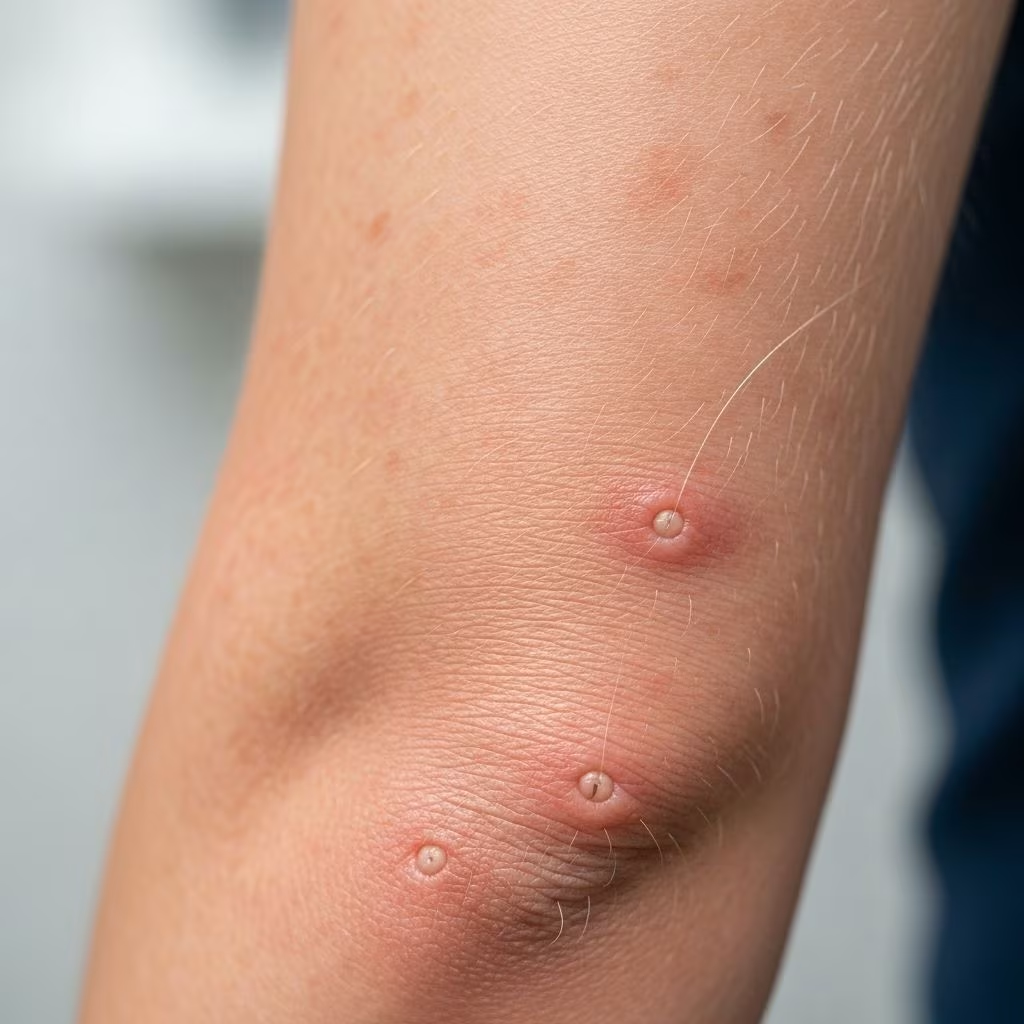
- Follicular papules: Small, rough bumps around hair follicles, giving the skin a sandpaper-like texture .
- Palmoplantar keratoderma: Marked thickening and yellow-orange discoloration of the palms and soles. Deep, painful fissures may develop .
- Islands of sparing: Distinct, unaffected skin patches interspersed within the rash—a classic diagnostic clue .
- Nail thickening: Often involving the whole nail, sometimes leading to discoloration and dystrophy .
- Hair loss (alopecia): Occasional, more common in severe or longstanding cases .
- Intense itching, burning, and skin tightness: These symptoms severely impact quality of life in active disease .
Severity may range from mild, localized plaques to diffuse erythroderma (redness and scaling over 90–100% of body surface area), sometimes accompanied by swelling of the legs and feet .
Table: Common Sites and Manifestations of PRP
| Region | Clinical Manifestations |
|---|---|
| Face & Scalp | Initial patches, scaling, redness |
| Trunk & Limbs | Spread of scaly plaques, follicular papules; sparing possible |
| Palms & Soles | Thickening, keratoderma, fissures |
| Nails | Thickening, slow growth, discoloration |
| Hair/scalp | Possible shedding, thinning |
Classification: Types of PRP
The most widely accepted classification is Griffiths’ six-type system, based on age at onset, clinical features, and chronicity :
- Type I: Classical Adult Type
The most prevalent form. Onset in adulthood with widespread plaques, palmoplantar keratoderma, and often spontaneous remission within several years. - Type II: Atypical Adult Type
Persistent, often generalized, with prominent ichthyosiform scaling and thickened skin. May persist for decades. - Type III: Classical Juvenile Type
Onset in childhood; similar features to classical adult type. Often self-limited, remission within a year is possible. - Type IV: Circumscribed Juvenile Type
Localized to elbows, knees, palms, and soles in children; usually resolves during adolescence. - Type V: Atypical Juvenile Type (Familial Form)
Early onset, persistent, sometimes inherited. Skin problems often last a lifetime . - Type VI: HIV-associated Type
Seen in HIV patients, often particularly severe and resistant to treatment.
Diagnostic Challenges and Procedures
Diagnosing PRP is challenging due to its rarity and similarity to other skin conditions:
- Clinical examination: Slower progression, islands of sparing, and follicular papules are important clues .
- Histopathology: A skin biopsy reveals thickening of the outer skin layers and characteristic changes in the hair follicles and keratin .
- Differential diagnosis: Important conditions to rule out include psoriasis, atopic dermatitis, lichenoid dermatoses, and pityriasis rosea .
- Genetic testing: May be considered in persistent, familial, or atypical juvenile cases to identify possible CARD14 gene mutations .
Differential Diagnosis
PRP’s clinical features overlap with other skin diseases, most notably psoriasis. Distinguishing features include:
- Islands of sparing are more characteristic of PRP than psoriasis.
- Follicular hyperkeratosis (rough papules) is more pronounced in PRP.
- Psoriasis tends to have thicker, silvery scales and does not usually cause palmoplantar keratoderma as severely as PRP.
- Response to psoriasis treatments is typically poor in PRP, helping to differentiate the two .
Other mimickers include eczema, lymphoma, drug eruption, and pityriasis rosea. Clinical expertise and biopsy are essential for accurate diagnosis.
Treatment Strategies
Management of PRP requires a tailored, stepwise approach combining topical and systemic therapies .
Topical Treatments
- Emollients and moisturizers: Alleviate dryness and soothe skin irritation.
- Topical corticosteroids: Useful for localized, mild cases—reduce inflammation and itching.
- Keratinolytics: Urea or salicylic acid may reduce thickening and scaling.
Systemic (Oral or Injectable) Treatments
- Retinoids (acitretin, isotretinoin): Highly effective for generalized PRP, promoting skin cell turnover .
- Methotrexate: Used in severe or refractory cases, especially when retinoids are unsuitable.
- Biologics (e.g., TNF-α inhibitors, IL-17 or IL-23 inhibitors): Increasingly used in PRP treatment, especially for those not responding to conventional therapies.
- Other immunosuppressive agents: Cyclosporine or azathioprine may be considered in resistant disease.
Adjunctive Measures
- Phototherapy: Narrowband UVB or PUVA, cautiously in selected patients.
- Wound and skin care: Prevent fissures and secondary infections.
- Psychological support: Chronic skin disease can cause significant distress; holistic care is important.
Treatment is often long-term and requires close monitoring for side effects. Responses vary significantly among individuals, and some cases remit spontaneously.
Prognosis and Long-term Care
- Classical adult PRP often eventually resolves within 3–5 years, but lingering symptoms may persist .
- Atypical or familial cases may have lifelong symptoms .
- Complications include skin fissures, thickening, infection risk, nail dystrophy, and rarely eye or oral complications .
- Quality of life can be severely affected, especially during periods of active, extensive disease.
Regular follow-up with a dermatologist is recommended to monitor disease activity, assess treatment efficacy, and manage side effects.
Living with PRP: Patient Guidance
- Adherence to therapy is crucial—do not stop medications without consulting a physician.
- Skin care routine: Use gentle cleansers, frequent moisturizers, and avoid irritants.
- Monitor for complications such as deep fissures, infections, and vision changes; seek timely medical advice.
- Seek support: Patient advocacy groups, counseling, and connecting with others may help cope with chronic symptoms.
Frequently Asked Questions (FAQs)
What triggers Pityriasis Rubra Pilaris?
The precise cause of PRP remains unknown in most cases. For familial or inherited forms, genetic mutations (such as CARD14) are implicated. No consistent environmental trigger is identified.
Can PRP be cured?
There is currently no permanent cure for PRP. Some forms, particularly classical adult and some juvenile types, can remit spontaneously. Treatment focuses on controlling and alleviating symptoms.
Is PRP life-threatening?
PRP is not generally life-threatening, but severe, generalized involvement can impair life quality and increase risk of secondary infections or complications, requiring close medical supervision.
What supportive therapies help people with PRP?
Supportive therapies include regular use of moisturizers, proper skin hygiene, psychological counseling, and monitoring for complications. Patient education and community support also play crucial roles.
How is PRP different from psoriasis?
Both diseases cause scaling plaques, but PRP often shows “islands of sparing” (normal skin amidst rash), pronounced follicular papules, and more severe palm and sole involvement. Histological examination and poor response to standard psoriasis treatments further distinguish PRP.
References
- https://www.ncbi.nlm.nih.gov/books/NBK482436/
- https://en.wikipedia.org/wiki/Pityriasis_rubra_pilaris
- https://rarediseases.org/rare-diseases/pityriasis-rubra-pilaris/
- https://www.healthline.com/health/pityriasis-rubra-pilaris
- https://medlineplus.gov/genetics/condition/familial-pityriasis-rubra-pilaris/
- https://dermnetnz.org/topics/pityriasis-rubra-pilaris
- https://www.msdmanuals.com/professional/dermatologic-disorders/psoriasis-and-scaling-diseases/pityriasis-rubra-pilaris
- https://rarediseases.info.nih.gov/diseases/7401/pityriasis-rubra-pilaris