

Recognizing and Treating Erythema Nodosum: Comprehensive Clinical Overview
Erythema nodosum (EN) is an inflammatory condition that affects the layer of fat beneath the skin, leading to the sudden development of tender, red or purple nodules. Most commonly seen on the shins, EN may herald underlying systemic illness, medication reactions, or infections, and warrants attentive recognition and management for optimal patient outcomes. This article provides an extensive overview of the disorder—its clinical features, etiology, diagnostic workup, and up-to-date treatment strategies.
Introduction
Erythema nodosum is best classified as a form of panniculitis, an inflammation of subcutaneous fat. The disorder presents as acute, firm, tender nodules—primarily on the anterior shins—and often signals a broader underlying medical process. Although it is typically self-limited, correct identification is crucial, as EN may require investigation for systemic disease or could arise from treatable secondary causes.
Clinical Features of Erythema Nodosum
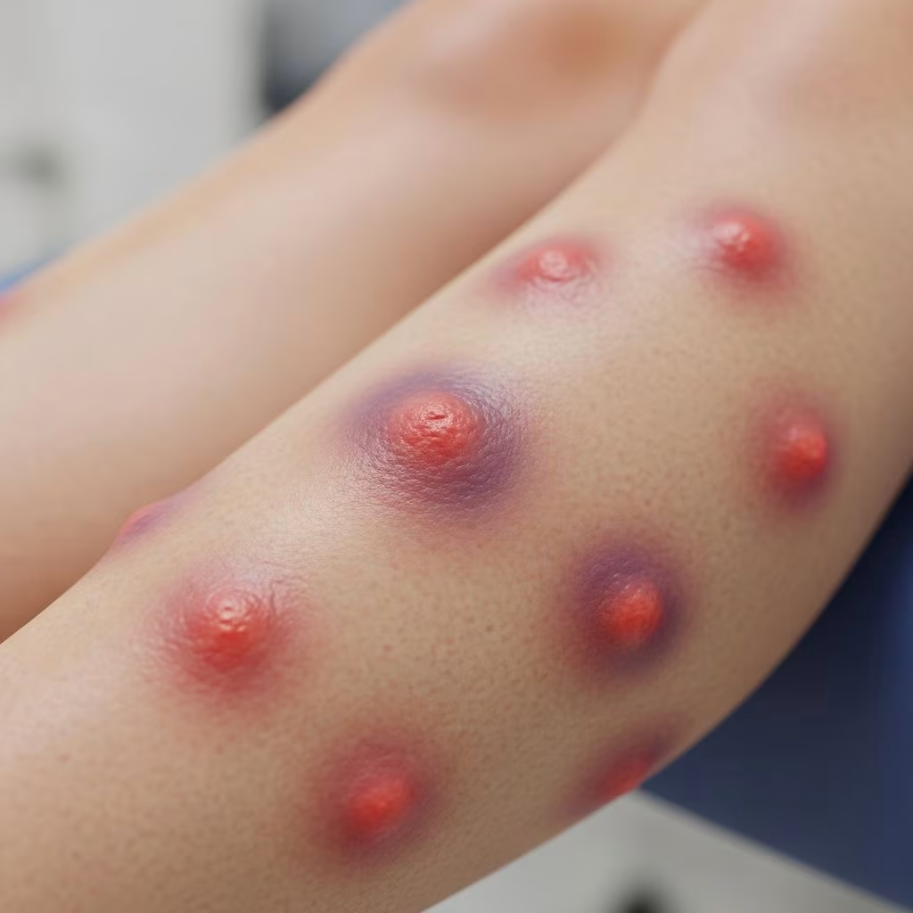
The hallmark of erythema nodosum is the presence of painful, red or violet lumps (nodules) under the skin. Important clinical characteristics include:
- Location: Bilateral anterior shins in the majority of cases, though lesions may also appear on forearms, thighs, trunk, and other extremities.
- Appearance: Nodules are round to oval, ranging from the diameter of a penny to a lime.
- Texture: Lesions are tender, warm, indurated (hard), and often feel deep to palpation.
- Color Evolution: Initially bright red, then progressing to bluish-purple or brown as healing ensues, eventually resembling bruises before fading.
- Resolution: Nodules resolve within 1-2 months, typically without scarring, though residual skin changes such as pitting may occur.
Additional systemic symptoms sometimes accompany the skin findings:
- Low-grade fever
- Malaise and fatigue
- Arthralgia (joint pain), especially knees and ankles
- Swollen lymph nodes
- Gastrointestinal symptoms: abdominal pain, diarrhea, vomiting (occasionally)
- Respiratory: cough, sore throat (if associated infection present)
Chronic erythema nodosum may rarely develop, with nodules merging into larger plaques, typically with less tenderness.
Epidemiology and Risk Factors
Erythema nodosum affects both adults and children, most commonly presenting in young adults between ages 20 and 40. Women are affected more than men, with up to a threefold higher prevalence among females. EN is not contagious, but its underlying causes may be infectious.
Key risk factors include:
- Recent streptococcal or other systemic infection
- Medication exposure (antibiotics, oral contraceptives)
- Systemic inflammatory diseases (sarcoidosis, IBD)
- Pregnancy, especially second trimester
- Genetic and regional factors (certain infections prevalent in geographic areas)
Etiology and Pathogenesis
EN is a reactive process—a skin manifestation provoked by antigenic stimuli. Its pathogenesis involves immune-mediated inflammation of subcutaneous fat (septal panniculitis), with a mixed cellular infiltrate but no vasculitis. About 30%–50% of cases are idiopathic (no clear cause), but identifiable triggers include:
Infectious Causes
- Bacterial: Most commonly group A streptococcal infection (pharyngitis, tonsillitis)
- Viral: Epstein-Barr (mononucleosis), hepatitis B and C, HIV
- Fungal: Coccidioidomycosis, histoplasmosis
- Parasitic: Giardia, amebiasis
Drug-Induced Causes
- Antibiotics: Penicillins, sulfonamides, amoxicillin
- Oral contraceptives and hormone therapies
- Miscellaneous: TNF-alpha inhibitors, bromides, iodides
Non-Infectious Medical Conditions
- Inflammatory Bowel Disease (IBD): Crohn’s disease, ulcerative colitis
- Sarcoidosis: Especially as part of Lofgren syndrome (EN, arthritis, hilar lymphadenopathy)
- Behcet disease, Sweet syndrome
- Malignancy: Leukemias and lymphomas
- Pregnancy: Hormonal changes thought to provoke immune reaction (often second trimester)
Underlying Conditions Associated with Erythema Nodosum
EN may be the first sign of an important systemic illness, sometimes preceding diagnosis of tuberculosis, deep fungal infections, sarcoidosis, or inflammatory bowel disease. Less commonly, it may signal an underlying malignancy such as lymphoma or leukemia.
| Category | Common Causes |
|---|---|
| Infection | Strep throat, TB, coccidioidomycosis, hepatitis, HIV |
| Medication | Penicillin, sulfa drugs, oral contraceptives |
| Autoimmune/Systemic Disease | Sarcoidosis, IBD, Behcet’s, Sweet’s syndrome |
| Pregnancy | Hormonal changes (second trimester) |
| Malignancy | Leukemia, lymphoma |
| Idiopathic | No identifiable cause (30%-50% cases) |
Diagnostic Approach
Diagnosis of EN is largely clinical, based on characteristic appearance and distribution of nodules. However, thorough evaluation for underlying disease is crucial.
Key Points in Diagnosis
- History: Recent infections (especially respiratory), medication use, systemic symptoms (fever, joint pain), travel/epidemiological risk, pregnancy
- Physical exam: Bilateral, tender, erythematous nodules on shins; may appear on other sites
- Labs: CBC, inflammatory markers (ESR, CRP), throat culture, antistreptolysin O titer (for strep), chest X-ray (for sarcoidosis, TB), stool studies (if GI symptoms), serology for relevant infections
- Skin biopsy: Rarely needed, but may confirm diagnosis; shows septal panniculitis without vasculitis
Example Diagnostic Workup
- Complete blood count (CBC)
- ESR and CRP for inflammation
- Throat swab if pharyngitis suspected
- Chest X-ray for sarcoidosis or tuberculosis
- Urine/blood/stool cultures if systemic symptoms present
- Pregnancy test if indicated
In about half of cases, no underlying trigger is found (idiopathic EN).
Differential Diagnosis
Other conditions that may mimic EN include:
- Cellulitis (diffuse soft tissue infection)
- Other types of panniculitis (lupus, Weber-Christian)
- Vasculitis (cutaneous polyarteritis nodosa)
- Fat necrosis
- Trauma-induced bruising
- Erythema induratum (related to tuberculosis)
Management and Treatment
EN is generally self-limited, resolving in 3–8 weeks as the immune reaction subsides. Treatment centers on supportive care and addressing underlying causative factors.
General Supportive Measures
- Rest and elevation of affected limbs
- Cool compresses to relieve pain
- Nonsteroidal anti-inflammatory drugs (NSAIDs) such as ibuprofen or naproxen for pain and inflammation
- Mild analgesics (acetaminophen)
- Compression stockings may reduce swelling
Treating Underlying Causes
- If infection identified: Specific antibiotics or antifungals as appropriate
- Discontinue causative medications (antibiotics, contraceptives)
- If associated with inflammatory disease: Management of underlying disease (IBD, sarcoidosis)
- Corticosteroids (oral or topical): Reserved for severe or resistant cases, but typically avoided unless underlying infection has been ruled out
When to Refer
- No improvement with conservative management within weeks
- Diagnostic uncertainty or atypical presentation
- Suspected systemic underlying illness needing specialist input
Complications and Prognosis
EN is commonly benign and self-limiting, usually leaving no permanent skin changes. Complications are uncommon, but include:
- Pigmentation changes (temporary)
- Persistent pain or discomfort
- Skin atrophy, pitting after resolution
- Recurrence, especially if underlying disease is not adequately treated (may be triggered by future pregnancy)
Most patients recover within 1–2 months. Chronic or recurrent EN may require further investigation.
Prevention and Patient Education
Prevention of EN depends on controlling triggers:
- Timely treatment of infections (especially strep)
- Cautious use of medications known to trigger EN
- Monitoring for recurrence in high-risk settings (e.g. pregnancy, ongoing IBD, sarcoidosis)
Patient education is essential regarding self-care measures, expected course, and warning signs indicating systemic involvement.
Frequently Asked Questions (FAQs)
Q: Is erythema nodosum contagious?
A: EN itself is not contagious. However, some underlying causes (such as strep throat or viral infections) may be infectious.
Q: Will the nodules leave scars?
A: Nodules typically heal without scarring, though some pitting or pigmentation changes may persist temporarily.
Q: How long does erythema nodosum last?
A: Most episodes resolve within 1–2 months, with gradual fading of lesions.
Q: Can erythema nodosum indicate a serious underlying illness?
A: Yes, EN can be a marker for systemic disease such as tuberculosis, sarcoidosis, or chronic infections. Full evaluation is important.
Q: What should I do during an episode?
A: Rest affected limbs, use pain relief measures (NSAIDs), and follow up with your health provider to rule out treatable causes or underlying illnesses.
Summary Table: Recognition and Management of Erythema Nodosum
| Feature | Details |
|---|---|
| Common Age Group | 20–40 years |
| Typical Lesions | Bilateral tender nodules on shins |
| Color Change | Red → purple → brown → fade |
| Key Causes | Strep infection, IBD, medication, sarcoidosis, pregnancy |
| Diagnosis | Clinical appearance, history, lab tests, skin biopsy (rare) |
| Primary Treatment | Supportive care, address underlying cause |
| Prognosis | Self-limiting, resolves in 1–2 months |
Conclusion
Recognizing erythema nodosum is a vital skill for clinicians and patients alike, as this seemingly simple skin disorder can be a window into broader systemic processes. Timely diagnosis, thorough evaluation for underlying causes, and appropriate supportive care constitute the cornerstones of successful treatment. Patients should be reassured about the benign and self-limited nature of most cases while encouraged to seek medical advice to exclude more serious triggers. Ongoing research continues to clarify the links between EN and systemic illnesses, underscoring the evolving importance of a multidisciplinary approach in its management.
References
- https://www.webmd.com/skin-problems-and-treatments/erythema-nodosum
- https://www.mountsinai.org/health-library/diseases-conditions/erythema-nodosum
- https://my.clevelandclinic.org/health/diseases/24565-erythema-nodosum
- https://www.ncbi.nlm.nih.gov/books/NBK470369/
- https://www.aafp.org/pubs/afp/issues/2007/0301/p695.html
- https://dermnetnz.org/topics/erythema-nodosum
- https://www.healthdirect.gov.au/erythema-nodosum