

Pancreatic diseases often require advanced diagnostic and therapeutic interventions. Two critical endoscopic techniques—Endoscopic Ultrasound (EUS) and Endoscopic Retrograde Cholangiopancreatography (ERCP)—have revolutionized the field. This guide offers an in-depth comparison of these procedures, their indications in pancreatic diagnostics, their respective strengths, and their ideal roles in modern clinical practice.
Introduction
The diagnosis and management of pancreatic and biliary diseases challenge gastroenterologists due to the deep anatomic location and complex physiology of these organs. Over the past decades, EUS and ERCP have become indispensable. EUS offers unparalleled imaging and tissue sampling, while ERCP provides both diagnostic imaging and critical therapeutic interventions.
Methods Overview
Endoscopic Ultrasound (EUS)
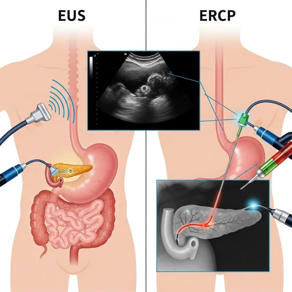
EUS combines endoscopy and high-frequency ultrasound to generate detailed images of the pancreas and surrounding structures. A thin, flexible endoscope equipped with an ultrasound probe is advanced through the patient’s mouth into the stomach and duodenum. The proximity of the probe to the pancreas ensures high-resolution imaging, facilitating the identification of small lesions and allowing for fine-needle aspiration (FNA) or biopsy if required.
Key features:
- High-resolution imaging of the pancreas, bile ducts, and surrounding tissues
- Ability to perform FNA/biopsy for cytologic or histopathologic diagnosis
- Useful for both diagnostics and certain therapeutic interventions
Endoscopic Retrograde Cholangiopancreatography (ERCP)
ERCP uses a side-viewing endoscope fitted with a camera to visualize the duodenum and access the biliary and pancreatic ducts. Dye is injected into these ducts under fluoroscopic (X-ray) guidance, allowing direct visualization of strictures, stones, or tumors. ERCP is primarily therapeutic, although it retains certain diagnostic capabilities such as tissue sampling and manometry.
Key features:
- Direct access to the biliary and pancreatic duct systems
- Therapeutic interventions (stone extraction, stenting, sphincterotomy)
- Less sensitive than EUS for parenchymal lesions outside the ductal system
Indications and Clinical Roles
Endoscopic Ultrasound (EUS)
- Characterization of pancreatic masses, cysts, and lesions
- Assessment and staging of pancreatic, gastroesophageal, and biliary cancers
- Evaluation of idiopathic acute and chronic pancreatitis
- Detection of microlithiasis and biliary sludge not seen on conventional imaging
- Guidance for interventional procedures (drainage, celiac plexus block, pseudocyst management)
Endoscopic Retrograde Cholangiopancreatography (ERCP)
- Treatment of biliary and pancreatic ductal obstruction (strictures, stones)
- Therapeutic stenting in malignant and benign ductal strictures
- Management of biliary leaks, fistulas, and papillary stenosis
- Acquisition of ductal tissue and fluid samples for cytology/biopsy
- Manometry of sphincter function
While both modalities have diagnostic implications, ERCP’s primary value lies in therapy, while EUS excels in diagnosis, staging, and tissue acquisition.
Diagnostic Performance and Accuracy
EUS is highly sensitive for detecting even small pancreatic lesions, cysts, and peri-pancreatic lymph nodes. Studies show EUS surpasses CT and MRI in identifying small (<2 cm) lesions and in diagnosing idiopathic acute pancreatitis (IAP) of biliary origin, such as microlithiasis or biliary sludge. EUS-guided biopsies allow for real-time targeting of suspicious findings.
ERCP provides information about the ductal anatomy and can reveal strictures, stones, and leaks. However, its utility for visualization outside the ductal system is limited. With ancillary techniques (such as intraductal ultrasound or cholangiopancreatoscopy), ERCP’s diagnostic power increases, particularly for ductal lesions and biliary strictures.
Therapeutic Capabilities and Limitations
| Therapeutic Intervention | EUS | ERCP |
|---|---|---|
| Biliary/Pancreatic Duct Stone Extraction | No | Yes |
| Stent Placement (biliary/pancreatic) | Emerging/limited | Yes |
| Pseudocyst Drainage | Yes | Limited |
| Celiac Plexus Block | Yes | No |
| Tumor Ablation/Injection | Yes | No |
| Ductal Tissue Sampling (Biopsy/Brushings) | Yes (FNA/FNB) | Yes (brush/forceps) |
While ERCP remains the gold standard for stone removal, stenting, and immediate management of ductal obstructions, EUS-guided therapies (drainage, injections, and tumor ablation) are rapidly evolving and increasingly used, especially when ERCP fails or is not feasible.
Safety Profile and Complications
EUS: Safety and Risks
- Sore throat, transient bloating, reactions to sedatives
- Rare: Perforation (stomach or duodenal wall)
- Risks with FNA: Bleeding, pancreatitis, infection (antibiotics often used for cyst puncture)
- Overall complication rate is lower than ERCP
ERCP: Safety and Risks
- Up to 7% overall complication rate
- Most common: Acute pancreatitis post-ERCP (1–5%)
- Bleeding, cholangitis, perforation, infection
- Risks increase with therapeutic interventions and in high-risk populations
EUS is generally safer than ERCP, especially when used for purely diagnostic purposes. ERCP, while highly effective, carries significant risks, especially for post-ERCP pancreatitis.
Integration of EUS and ERCP
There is a trend towards integrating the diagnostic precision of EUS with the therapeutic power of ERCP. The “EURCP” model involves performing both procedures in a single session, maximizing diagnostic yield and facilitating immediate therapy when needed. This integrated approach improves patient outcomes, reduces hospital visits, and can be more cost-effective, provided the clinical setting and endoscopist expertise are appropriate.
- Benefits:
- One sedation, less patient movement
- Immediate transition from diagnosis to therapy
- Potential avoidance of other imaging and delayed diagnoses
- Drawbacks:
- Longer procedure and sedation times
- Requires a high level of expertise
- Not appropriate for all patient populations
Clinical Decision-Making: When to Use Which?
- Suspected pancreatic mass, cyst, or need for tissue diagnosis: Prioritize EUS for its sensitivity and ability to biopsy; only follow with ERCP if therapy is needed.
- Obstructive jaundice with evidence of biliary stricture or stone: Begin with EUS for diagnosis and staging when malignancy is suspected. If immediate therapy is required, ERCP for stone removal or stenting should be considered.
- Indeterminate biliary or pancreatic ductal strictures: Use EUS for imaging and biopsy, ERCP for ductal sampling and manometry, or both in combination when appropriate.
- Idiopathic acute pancreatitis: Use EUS for detection of microlithiasis or occult causes not visible on CT/MRI.
Future Directions and Innovations
The boundaries between diagnostics and therapy are blurring as EUS continues to evolve. Emerging EUS-guided procedures include biliary drainage (especially when ERCP fails), celiac plexus neurolysis, localized tumor therapies, and even direct intervention in previously inaccessible areas. Hybrid “EURCP” scopes and robotic assistance may further expand the capabilities and safety of these techniques in the coming years.
Comparison Table: EUS vs. ERCP
| Feature | Endoscopic Ultrasound (EUS) | Endoscopic Retrograde Cholangiopancreatography (ERCP) |
|---|---|---|
| Main Purpose | Diagnosis (Imaging, Tissue Sampling) | Therapy (primarily), plus limited diagnosis |
| Imaging Modality | High-frequency ultrasound | Fluoroscopy/X-ray |
| Ductal Access | Indirect (visualizes ducts + parenchyma) | Direct |
| Biopsy Capability | Yes (FNA/FNB under real-time guidance) | Yes (Brush, forceps, but limited) |
| Stone Management | No | Yes (extraction, lithotripsy) |
| Stenting | Emerging for select indications | Mainstay for ductal strictures |
| Complication Rate | Low (except with FNA/CNA) | Moderate; highest is post-ERCP pancreatitis |
| Role in Cancer | Diagnosis, staging, biopsy | Diagnosis (ducts), palliative stenting |
| First-Line for Idiopathic Acute Pancreatitis | Yes | No |
| Guidance for Therapy | Yes (emerging) | Yes |
Frequently Asked Questions (FAQs)
What is the main difference between EUS and ERCP?
EUS provides high-resolution, ultrasound-based imaging to visualize the pancreas and surrounding structures, while ERCP enables direct access and therapy of the bile and pancreatic ducts using X-ray guidance.
Which procedure is safer?
Diagnostic EUS generally has a lower complication rate than ERCP. ERCP complications include pancreatitis, bleeding, and infection, especially when therapeutic interventions are performed.
Can EUS replace ERCP?
No. EUS and ERCP are complementary. EUS excels at diagnosis and targeted tissue sampling, while ERCP remains the mainstay for ductal interventions such as stone extraction and stenting.
Are both procedures performed together?
Yes. In certain complex or unclear cases, both EUS and ERCP can be performed successively, offering immediate transition from diagnosis to therapy (the “EURCP” approach).
Is sedation required for both?
Yes, both EUS and ERCP are usually performed under moderate to deep sedation or general anesthesia for patient comfort and safety.
What emerging technologies are on the horizon?
Hybrid endoscopes, enhanced EUS-guided therapies (including biliary drainage), and real-time image fusion techniques are being developed to improve safety, diagnostic power, and therapeutic efficiency.
References
- https://ercpmalpractice.com/ercp-alternatives/endoscopic-ultrasound-vs-ercp/
- https://pmc.ncbi.nlm.nih.gov/articles/PMC10606726/
- https://pmc.ncbi.nlm.nih.gov/articles/PMC9745610/
- https://practicalgastro.com/2025/06/26/role-of-eus-and-ercp-in-diagnosis-and-management-of-idiopathic-pancreatitis/
- https://www.researchhub.blog/eus-vs-ercp-procedure-5-key-facts
- https://sciencespark.blog/eus-vs-ercp-procedure-guide