


Introduction
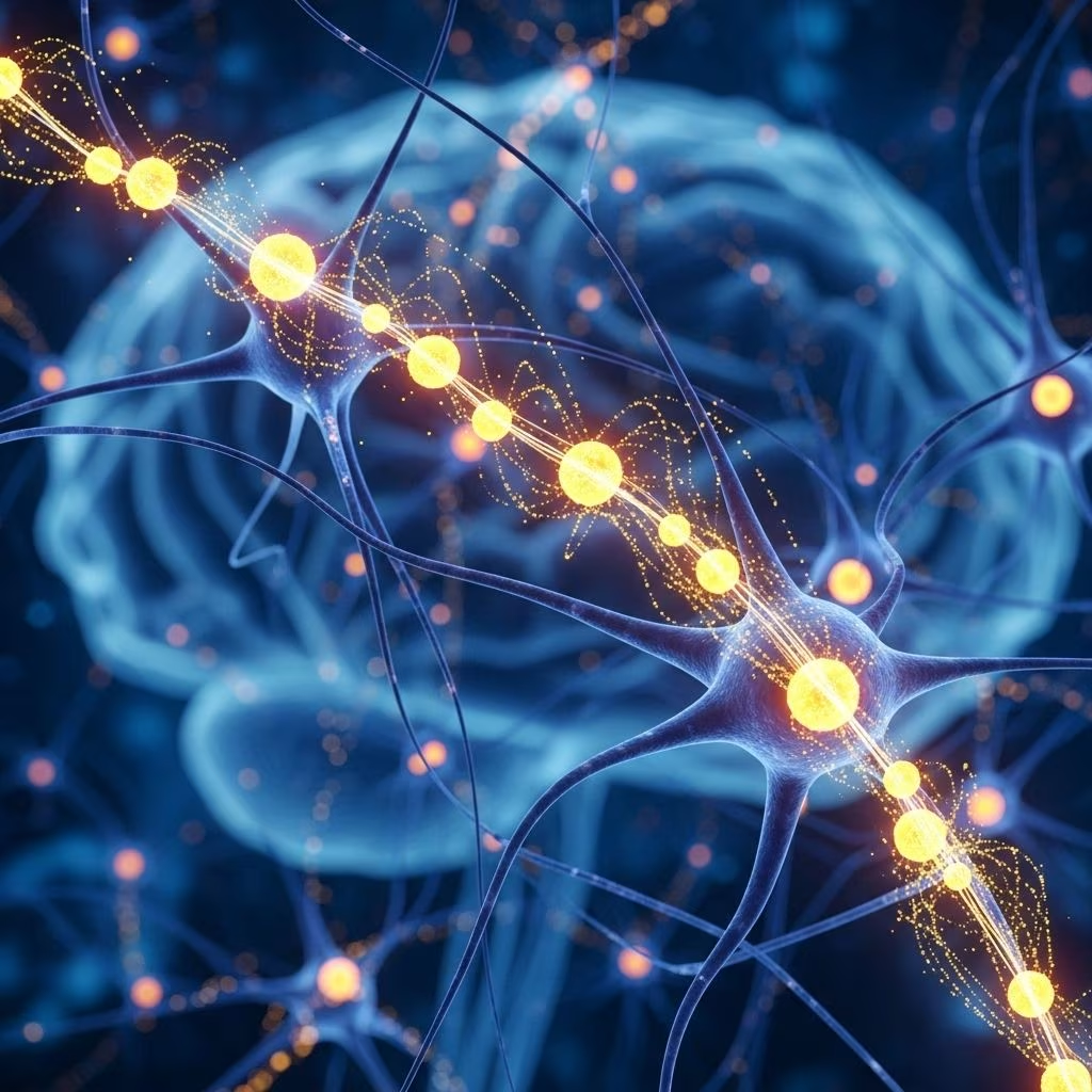
Dopamine plays a fundamental role in brain function, modulation of emotion, motivation, attention, and reward learning. Advances in neuroscience have allowed researchers to visualize dopamine release in real-time, employing cutting-edge imaging and biosensor technologies. The emergence of visualization videos, derived from complex neuroimaging datasets, now provides new insights into the spatiotemporal dynamics of dopamine signaling. This article explores the intersection of visualization approaches and dopamine release studies, highlighting technologies, biological mechanisms, research applications, and implications for understanding brain activity.
Understanding Dopamine: The Neurotransmitter and Its Role
Dopamine is a catecholamine neurotransmitter critically involved in regulating movement, reward perception, mood, and various cognitive processes. Dopaminergic neurons are primarily located in brain regions such as the ventral tegmental area (VTA) and the substantia nigra, projecting to networks that underpin both essential functions and complex behaviors.
- Dopamine is synthesized from the amino acid tyrosine.
- It influences pleasure, motivation, motor control, and reinforcement learning.
- Dysregulation in dopamine dynamics is linked to disorders like Parkinson’s disease, schizophrenia, and addiction.
The Biological Mechanism of Dopamine Release
Dopamine is released from presynaptic neurons into the synaptic cleft, where it binds to postsynaptic receptors to exert its effects. The process is tightly controlled by action potentials, calcium signaling, and specialized vesicular release machinery.
- Triggered by electrical impulses and calcium influx.
- Release occurs at specialized active zones, involving SNARE proteins like synaptobrevin-2 and scaffolding proteins such as Bassoon.
- Dopamine signaling is terminated by uptake via transporter proteins and enzymatic degradation.
Dendritic release of dopamine exhibits unique features, such as rapid and calcium-mediated efflux, allowing broad modulatory effects beyond classical synaptic transmission.
Visualization Technologies for Dopamine Release
Direct visualization of dopamine activity in the intact brain has become possible with the development of advanced imaging modalities. These techniques enable researchers to observe spatial and temporal patterns of dopamine release following pharmacological or behavioral stimulation.
- Positron Emission Tomography (PET)
- Electrochemical biosensors
- Composite nanofilms (DopaFilm)
- Optical imaging
These platforms generate large multidimensional datasets, which are then transformed into visualization videos, providing both quantitative and qualitative insights into neurotransmitter dynamics.
PET Imaging and Dopamine Visualization
Positron Emission Tomography is a gold-standard modality for studying endogenous dopamine fluctuations in the human brain. By administering radiolabeled tracers like [11C]-raclopride, PET scans track dopamine concentrations with high spatial and temporal resolution.
- Dynamic PET imaging generates 4D datasets, encoding dopamine concentration across space and time.
- Visualization options include:
- Cine loop videos: Animated slices showing dopamine changes over time.
- Peak-time parametric images: Static images summarizing the timing of dopamine peaks at each voxel location.
- Researchers validate these visualizations by exposing subjects to repeated stimuli and comparing spatio-temporal patterns of dopamine release.
These visualization videos have revolutionized the understanding of neurotransmitter timing and response dynamics, illuminating new forms of functional brain imaging beyond traditional anatomical studies.
The Use of 2D Composite Nanofilms in Visualizing Dopamine
Recent technological breakthroughs have introduced chemi-sensitive 2D composite nanofilms, such as DopaFilm, for the direct visualization of dopamine efflux at the synaptic level.
- DopaFilm enables detection of dopamine release with quantal sensitivity in real-time.
- The film covers a large spatial domain, capable of identifying dopamine release from both axons and dendrites.
- Fluorescence videos reveal hotspots corresponding to active zones and track the spread of dopamine in peri-synaptic and extra-synaptic spaces.
- Super-resolution imaging links anatomical protein markers to observed release activity, clarifying the molecular machinery involved.
Advantages:
- Unprecedented spatio-temporal resolution of dopamine signaling.
- Ability to distinguish release sites and diffusion patterns.
- Versatility for studying somatodendritic release mechanisms.
Visualization videos produced with nanofilm techniques provide unprecedented clarity into the dynamic nature of dopamine transmission, opening new avenues for neurobiological exploration.
Animal Models and Dopamine Monitoring
Animal models, particularly rodents, are widely used for direct study of dopamine release dynamics. Techniques such as electrical stimulation of dopaminergic neurons and carbon microelectrode measurements allow precise monitoring.
- Ventral tegmental area (VTA) neurons are stimulated to release dopamine into the nucleus accumbens (NAc), a key reward center.
- Electrical pulses trigger release, measurable via changes in electrical signals detected by microelectrodes.
- Pharmacological manipulation (administration of activators or inhibitors) alters dopamine release dynamics in situ.
Visualization of these recordings and videos offers researchers the ability to study cause-effect relationships, underlying network wiring, and the role of dopamine in animal behavior.
Viewing Visualization Videos: Scientific and Psychological Impacts
Visualization videos—created from imaging technologies and biosensor output—are much more than artistic representations. They offer researchers and students a means to directly observe and comprehend neurophysiological mechanisms in real time.
- Educational value: Videos demystify complex datasets, providing intuitive understanding of neurochemical events.
- Research utility: Visualization enables identification of activation patterns, release hotspots, and temporal changes not apparent in static images.
- Enhanced engagement: Videos improve retention and interpretation of data, helping bridge gaps between theory and empirical evidence.
- Facilitate hypothesis generation: Visual context sparks new questions about brain function, disease, and therapy.
By transforming abstract numbers into visible, evolving shapes and colors, visualization videos make palpable the dynamic processes occurring within living neural tissue.
Correlation Between Visualization Videos and Dopamine Dynamics
The process of viewing visualization videos—for both researchers and participants—has sparked interest in the possibility that observing dynamic representations of dopamine signals may itself influence dopamine-related brain processes. Several hypotheses and preliminary findings need to be considered:
- Mimetic effects: Viewing videos of dopamine activity may evoke reward-system responses, as the brain recognizes representations of pleasure and motivation.
- Neurofeedback: In some experimental protocols, subjects receive real-time visual feedback of their own dopamine activity, potentially affecting learning and self-regulation.
- Empathy and self-projection: Observers may mentally simulate the experiences depicted in visualization videos, leading to measurable neural activation.
Current evidence suggests that while viewing such videos offers invaluable scientific insights, the act of viewing does not directly trigger measurable changes in brain dopamine release without appropriate behavioral or pharmacological stimulus. However, the interaction between video content and observer perception remains a fertile area for further investigation.
Applications and Implications of Dopamine Visualization
The ability to visualize dopamine release is not merely a technical triumph—it has wide-reaching applications and profound implications for neuroscience, medicine, and beyond.
- Understanding psychiatric and neurological diseases: Visualization videos illuminate abnormalities in dopamine signaling, aiding diagnosis and treatment planning for conditions like Parkinson’s disease, bipolar disorder, ADHD, and addiction.
- Drug development: Researchers track the effects of experimental therapies in vivo, improving the efficiency of drug discovery.
- Educational platforms: Animated visualizations bring neuroscience into classrooms and public outreach, enhancing awareness and literacy.
- Neurofeedback therapy: Real-time visualization may facilitate brain-training protocols targeting attention, impulse control, or mood regulation.
| Method | Spatial Resolution | Temporal Resolution | Special Strengths |
|---|---|---|---|
| PET Imaging | High (voxel-level) | Moderate (seconds to minutes) | Quantitative mapping in humans |
| 2D Nanofilm (DopaFilm) | Super-resolution (synaptic level) | High (real-time) | Precise spatial and temporal release detection |
| Electrochemical Microelectrodes | Focal (single site) | Very high (milliseconds) | Direct measurement, animal models |
Limitations and Challenges in Dopamine Visualization
Despite the dramatic progress in visualizing dopamine release, several limitations remain:
- Spatial constraints: Human neuroimaging is generally coarser than animal or in vitro systems.
- Temporal limitations: Some imaging technologies lack millisecond-level time resolution, limiting analysis of fast neurotransmitter events.
- Probe specificity and sensitivity: Ensuring that sensors accurately reflect dopamine activity without cross-reactivity remains a technical challenge.
- Interpreting complex data: Visualization tools generate vast datasets that require advanced computational analysis and expertise.
- Cost and accessibility: Advanced imaging is expensive and not widely available outside research centers.
Continued research in sensor design, computational modeling, and collaborative standardization aims to address these obstacles and democratize access to visualization technologies.
Future Directions in Dopaminergic Visualization
The field is rapidly evolving, with several promising avenues:
- Integration of multimodal imaging platforms to combine anatomical, functional, and molecular data.
- Development of non-invasive, wearable biosensors for real-world dopamine monitoring.
- Application of artificial intelligence to automate detection and interpretation of release patterns in videos.
- Advancement of educational and neurofeedback tools using live dopamine visualization for training and treatment.
- Expansion into other neurotransmitters and molecular signaling pathways using visualization video technologies.
As visualization techniques move from bench to bedside, they hold the potential to transform patient care, scientific understanding, and societal conversations about brain and behavior.
Frequently Asked Questions (FAQs)
- Q: Can watching visualization videos of dopamine release stimulate my own dopamine production?
A: Simply watching such videos does not directly stimulate dopamine release in the brain, unless the content itself provides rewarding or motivational stimuli. The visualizations represent real data from experiments, but viewing them does not directly modify your neurochemistry.
- Q: What are the most advanced techniques for visualizing dopamine release?
A: PET imaging and 2D composite nanofilms (such as DopaFilm) currently represent the highest standard for spatial and temporal mapping of dopamine release in living brain tissue.
- Q: Why is dopamine visualization important for understanding diseases?
A: Abnormal dopamine signaling underlies several disorders. Visualization videos reveal where and when such abnormalities occur, guiding research and therapeutics.
- Q: Are these visualization techniques available outside research labs?
A: While technologies like PET are clinically available for some diagnostic purposes, most advanced visualization platforms remain limited to research settings due to cost and complexity.
References
- https://pmc.ncbi.nlm.nih.gov/articles/PMC3725608/
- https://elifesciences.org/articles/78773
- https://www.jove.com/v/31209/monitoring-dopamine-release-rat-model-with-vta-dopaminergic-neuron
- https://www.youtube.com/watch?v=6J73tzP2zFg
- https://www.youtube.com/watch?v=Wa8_nLwQIpg
- https://www.youtube.com/watch?v=uE2mRzDOXyA
- https://www.shutterstock.com/video/search/brain-dopamine
- https://www.youtube.com/watch?v=f7E0mTJQ2KM
- https://www.youtube.com/watch?v=QmOF0crdyRU