

Eosinophilic Leukemia Overview
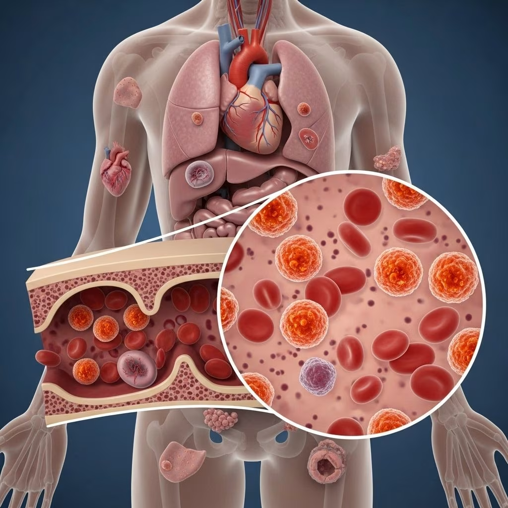
Eosinophilic leukemia is a rare cancer of the blood characterized by an abnormally high number of eosinophils—specialized white blood cells—in the blood, bone marrow, and sometimes other tissues. These cells play crucial roles in the immune system, primarily in combating parasitic infections and mediating allergic responses. When their production becomes dysregulated due to malignancy, the resulting condition is referred to as eosinophilic leukemia.
Leukemia encompasses a diverse group of cancers affecting blood cells. According to the National Cancer Institute, there were around 61,090 new leukemia diagnoses in the United States in 2021, representing 3.2% of new cancer cases. Eosinophilic leukemia is a subtype within chronic myelogenous leukemias and is far less common than other forms of leukemia. This article will detail the condition’s symptoms, risk factors, causes, diagnosis, treatment, and outlook, as well as provide answers to frequently asked questions.
Types of Leukemia and the Role of Eosinophils
- Acute vs. Chronic: Leukemia is classified by the speed of progression—acute forms grow quickly, while chronic types develop more slowly.
- Myeloid vs. Lymphoid: Classification also depends on which blood cell lineage is affected. Eosinophilic leukemia falls under the myeloid group, affecting cells derived from myeloblasts.
- Eosinophils: These white blood cells typically comprise less than 5% of the total white blood cell count. In leukemia, eosinophils exceed this limit and can damage organs through the release of toxic substances.
What Is Eosinophilic Leukemia?
Eosinophilic leukemia occurs when too many eosinophils are produced by the body in response to a blood cancer. The overload of these cells in blood, bone marrow, and tissues can harm organs. Eosinophilia is defined as a count of eosinophils greater than 500 per cubic millimeter (mm3) in a standard complete blood count; normal counts are 350 to 500 mm3.
There are two primary subtypes of eosinophilic leukemia:
- Chronic Eosinophilic Leukemia (CEL): A chronic myeloproliferative disorder driven by clonal expansion of eosinophilic precursors.
- Acute Eosinophilic Leukemia: A more rapidly progressive variant, with extensive immature eosinophil proliferation.
Why Are High Levels of Eosinophils Harmful?
Eosinophils can release chemicals such as cytokines and enzymes that cause inflammation and tissue damage. Persistent eosinophilia can lead to:
- Damage to heart, lungs, gastrointestinal tract, nervous system, and skin
- Blood clot formation and increased risk of organ failure
Symptoms of Eosinophilic Leukemia
The symptoms of eosinophilic leukemia are diverse and may resemble other illnesses. Common signs include:
- Fatigue
- Unexplained fever
- Swollen lymph nodes
- Night sweats
- Shortness of breath and cough
- Muscle pain and weakness
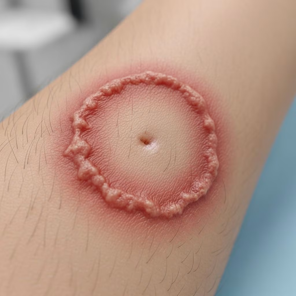
- Skin rashes (eczema, urticaria, angioedema, mucosal ulcers)
- Rhinitis (runny nose)
- Unintentional weight loss
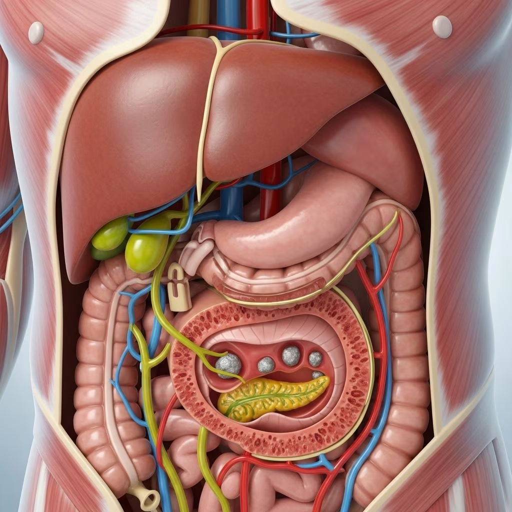
- Abdominal pain (if spleen or liver is enlarged)
- Bleeding or bruising easily
Complications depend on which organs are most affected. Sustained high levels of eosinophils may cause progressive heart failure, lung fibrosis, gastrointestinal issues, hypertension, neurological problems, and blood clots. In rare cases, skin or nail disorders may be the first sign.
Risk Factors
While most cases of eosinophilic leukemia are idiopathic (with no identifiable cause), a minority are linked to genetic abnormalities. Known risk factors include:
- Genetic mutations: Chromosomal abnormalities such as the FIP1L1-PDGFRA fusion gene. This abnormality may predispose individuals to uncontrolled eosinophil production.
- Family history: Rarely, a hereditary pattern of leukemia or eosinophilia exists.
- Pre-existing blood disorders: Some individuals with chronic myeloproliferative diseases may be at increased risk.
- Exposure to certain chemicals: While most cases aren’t linked to toxins, some research indicates potential contributions from pesticides or industrial agents.
Because these risk factors are uncommon and the genetic basis is often unclear, preventive measures are not well established for eosinophilic leukemia.
Causes and Disease Mechanism
| Cause/Mechanism | Description |
|---|---|
| Genetic mutation | Mutations such as FIP1L1-PDGFRA cause unregulated proliferation of eosinophils |
| Idiopathic origin | Most cases have no identifiable origin; termed idiopathic |
| Abnormal cell development | Eosinophilic leukemia arises when myeloid stem cells in bone marrow undergo malignant transformation at the blast (immature) stage |
The underlying disease process involves uncontrolled clonal proliferation of abnormal eosinophils. These immature cells accumulate in the bone marrow and peripheral blood.
Diagnosis of Eosinophilic Leukemia
Accurate diagnosis is essential for prognosis and treatment planning. Diagnostic steps usually include:
- Medical history and physical exam: Assessing symptoms such as fever, lymph node swelling, rash, and organ dysfunction.
- Complete blood count (CBC): A key test showing elevated eosinophils (>500/mm3).
- Bone marrow biopsy: Enables direct visualization of abnormal eosinophil proliferation.
- Cytogenetic and molecular studies: Detect chromosomal abnormalities such as FIP1L1-PDGFRA fusion.
- Imaging studies: Ultrasound, CT, or MRI may be used to identify organ enlargement or damage.
Diagnosis may require ruling out secondary causes of eosinophilia, such as parasitic infections, allergies, drug reactions, or other cancers. Physicians often differentiate eosinophilic leukemia from hypereosinophilic syndrome (HES), which shares overlapping features but may not involve a malignant process.
Treatment of Eosinophilic Leukemia
Timely treatment is essential to control symptoms and prevent organ damage. The main treatment options include:
- Chemotherapy: Standard regimens suppress the abnormal proliferation of eosinophils. Drugs may include hydroxyurea.
- Steroids: Systemic corticosteroids reduce inflammation and lower eosinophil counts rapidly.
- Tyrosine kinase inhibitors (TKIs): Targeted drugs such as imatinib are highly effective in cases involving FIP1L1-PDGFRA mutation.
- Immunotherapy: Sometimes used as an adjunct for resistant cases.
- Radiation therapy: Used mainly for palliative purposes if organs are compromised.
- Supportive care: Managing symptoms, preventing complications such as infections or blood clots, and monitoring cardiac and renal function.
The choice of therapy depends on clinical presentation and genetic findings. Some cases respond remarkably well to targeted therapy, especially when a known mutation is present. Surgery is rarely used, except for biopsy or when organ swelling threatens function.
Outlook and Prognosis
The prognosis for eosinophilic leukemia is highly variable, depending on:
- Subtype of leukemia (chronic vs. acute)
- Severity and presence of organ damage
- Age and overall health status
- Genetic markers (predicting response to targeted therapy)
Morbidity and mortality are mainly due to organ damage—heart failure and blood clots are common lethal complications. CEL-NOS (chronic eosinophilic leukemia, not otherwise specified) can progress to acute myelogenous leukemia, which is aggressive and life-threatening if untreated.
Early detection and prompt, personalized therapy can improve outcomes and long-term survival. Regular follow-up is crucial, as relapse or transformation is possible.
Living with Eosinophilic Leukemia
Management beyond medical therapy involves patient education, symptom monitoring, and supportive care. Individuals are encouraged to:
- Maintain regular contact with healthcare providers
- Understand and recognize warning signs of relapse
- Adhere to prescribed medications and attend follow-up appointments
- Engage in healthy lifestyle practices (balanced diet, moderate exercise, avoiding infections)
Psychological support, access to counseling, and involvement in patient advocacy groups can be valuable resources.
Frequently Asked Questions (FAQs)
Q: Is eosinophilic leukemia the same as hypereosinophilic syndrome (HES)?
A: No, while they share similar features (high eosinophil counts), eosinophilic leukemia is malignant, with clonal expansion of abnormal cells, whereas HES may not involve cancerous changes. Molecular studies help distinguish the two.
Q: What causes the increase in eosinophils in this disease?
A: In most cases, the cause is unknown. Sometimes, genetic abnormalities such as the FIP1L1-PDGFRA fusion gene are responsible for unregulated eosinophil production.
Q: Who is at risk for eosinophilic leukemia?
A: The risk is highest in individuals with certain genetic mutations or pre-existing blood disorders, but most cases are idiopathic and unpredictable.
Q: How is eosinophilic leukemia treated?
A: Treatment may include chemotherapy, steroids, targeted tyrosine kinase inhibitors, and supportive care. The exact regimen is individualized based on genetic findings and organ involvement.
Q: Can eosinophilic leukemia be prevented?
A: There are no proven preventive measures due to the unclear risk factors and genetic origins for most cases.
Q: What is the long-term outlook for someone diagnosed with eosinophilic leukemia?
A: Outlook depends on early detection, effective treatment, subtype, and presence of organ damage. Some patients achieve remission, while others may experience recurrent disease or severe complications.
Resources for Patients and Caregivers
- National Cancer Institute: Current research, statistics, and support resources for leukemia patients.
- Leukemia & Lymphoma Society: Patient advocacy, support groups, financial assistance, and educational resources.
- Medical Genetics Clinics: Testing and counseling for families concerned about hereditary risk.
- Online platforms: Newsletters and forums for sharing experiences and accessing peer support.
Key Takeaways
- Eosinophilic leukemia is a rare blood cancer, distinguished by an excess of eosinophils resulting in potential organ damage.
- Symptoms are non-specific and include fatigue, fever, lymphadenopathy, rashes, and signs of organ dysfunction.
- Diagnosis requires blood tests, bone marrow biopsy, and genetic studies.
- Treatment varies by individual and includes chemotherapy, steroids, and targeted therapy.
- Prognosis is improved with early intervention and proper management but may remain guarded in advanced cases involving organ damage.
References
- https://www.ebsco.com/research-starters/health-and-medicine/eosinophilic-leukemia
- https://www.healthline.com/health/leukemia/eosinophilic-leukemia
- https://pmc.ncbi.nlm.nih.gov/articles/PMC4966405/
- https://www.medicalnewstoday.com/articles/what-is-chronic-eosinophilic-leukemia
- https://www.healthline.com/health/eosinophil-count-absolute
- https://medlineplus.gov/genetics/condition/pdgfrb-associated-chronic-eosinophilic-leukemia/