

Lattice degeneration is a relatively common retinal condition that affects the peripheral retina and may increase the risk of serious complications, including retinal tears and detachments. While not everyone with lattice degeneration will experience vision problems, understanding this condition is crucial for maintaining long-term retinal health. This guide covers everything you should know about lattice degeneration in your eyes—including its definition, symptoms, causes, risks, available treatments, prevention, and answers to frequently asked questions.
About Lattice Degeneration
Lattice degeneration is characterized by a gradual thinning of the retina, mainly in its peripheral regions—that is, the areas farther from the center of vision. This particular pattern of degeneration affects roughly 6–10% of the general population, with estimates sometimes reaching as high as 10% depending on the diagnostic criteria and methods used. It most often occurs in both eyes, with about one-third to one-half of affected individuals experiencing bilateral involvement.
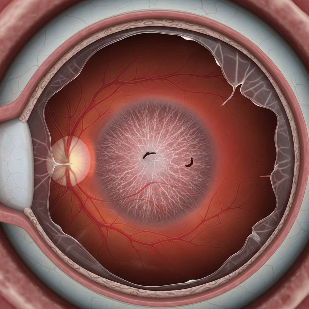
The retina itself is a thin, sensitive tissue lining the back of your eye, responsible for capturing light and sending electrical signals to the brain, allowing you to see. Any condition affecting the retina can potentially threaten visual function. Lattice degeneration, specifically, leads to localized patches where this tissue thins, sometimes forming cigar-shaped or oval lesions with distinct appearances during eye examinations.
- The peripheral retina, where lattice degeneration develops, is crucial for side (peripheral) vision and night vision.
- The macula—the central portion of the retina—primarily controls acuity (sharpness) and color perception and is typically not affected by lattice degeneration.
- Lesions caused by lattice degeneration may display a cross-hatching pattern from whitened or sclerotic retinal vessels, or clusters of pigment, and they may be present as a single or multiple patches in each eye.
Symptoms of Lattice Degeneration
Many people with lattice degeneration are completely asymptomatic, meaning they do not experience noticeable vision problems or warning signs. In most cases, lattice degeneration is discovered during a routine, dilated eye exam performed by an eye care professional.
Possible Signs and Symptoms
- Lattice degeneration itself often does not directly cause vision loss or significant visual symptoms.
- Most commonly discovered incidentally during routine examinations.
- If complications such as a retinal tear or retinal detachment occur, then symptoms may become pronounced, including:
- Sudden appearance or increase in floaters (tiny specks or strings drifting through your field of vision)
- Flashes of light in the peripheral vision
- Sudden shadow or curtain over part of the visual field
- Loss of peripheral vision
If you experience any of the above symptoms, especially a sudden increase in floaters or flashes, or notice a shadow over your vision, seek immediate medical attention, as these could be signs of a retinal tear or detachment—a sight-threatening emergency.
Causes and Risk Factors of Lattice Degeneration
The precise cause of lattice degeneration remains unclear. The condition seems to arise from a combination of local retinal thinning, changes in overlying vitreous (the gel-like substance filling the eye), and areas of tightly adherent vitreoretinal interface.
Key Risk Factors
- Myopia (Nearsightedness): Lattice degeneration is more common in people with myopia, particularly those with moderate degrees of nearsightedness. Studies suggest a prevalence as high as 33% in myopic eyes, compared to an average of 6–10% in the general population.
- Hereditary influences: Certain genetic syndromes—such as Stickler and Wagner syndromes—may predispose individuals to lattice degeneration, typically by affecting the structure of connective tissues in the eye.
- Age: Lattice degeneration is often discovered in young adults but can continue to progress with age.
- Family history: There may be a hereditary component for those with close relatives affected by the condition.
- History of retinal detachment: People who have had a retinal detachment—or have a family history of one—are at increased risk for lattice degeneration as well as future detachments.
What Causes Lattice Lesions?
Though the mechanisms are not fully understood, possible contributing factors include local vascular insufficiency (decreased blood flow), vitreous traction (pulling on the retina by the vitreous), and degeneration of retinal cells due to genetic factors, aging or other metabolic factors.
Does Lattice Degeneration Lead to Blindness?
Lattice degeneration itself rarely causes vision loss or blindness directly; however, it is a recognized risk factor for retinal tears and rhegmatogenous retinal detachment. These complications, if left untreated, can result in permanent vision loss or blindness.
How Common is Retinal Detachment in Lattice Degeneration?
- Up to 30% of retinal detachments are associated with sites of lattice degeneration.
- However, the vast majority of people with lattice degeneration will never experience a retinal detachment; estimates suggest retinal detachment develops in about 0.7% of eyes with lattice degeneration.
Most cases remain stable over time; only a minority require intervention, and vision-threatening complications are rare if the condition is monitored and managed appropriately.
Treatment Options for Lattice Degeneration
There is no specific cure for lattice degeneration, and most people do not require active treatment. Instead, regular monitoring and patient education form the cornerstone of management. Treatment is considered primarily in cases where there are high-risk features or symptomatic complications.
Monitoring
- The majority of patients should have annual dilated eye exams to monitor for retinal changes, new lesions, or evidence of complications.
- People with lattice degeneration should be informed about the symptoms of retinal tears or detachment (e.g., sudden floaters, flashes, loss of vision) so that they can seek prompt care if such events occur.
When Is Treatment Needed?
- Treatment for lattice degeneration itself is not routinely recommended unless there is an associated retinal tear, a history of retinal detachment, or if other risk factors are present.
- In select cases, an eye doctor may recommend laser photocoagulation (laser therapy) or cryotherapy (freezing treatment) to strengthen the thinned retina and reduce the risk of progression to a tear or detachment.
- The effectiveness of prophylactic (preventive) treatment to reduce the risk of retinal detachment is debated, with studies showing mixed results on its value for preventing detachments in people with asymptomatic lattice degeneration.
Treatment for Retinal Tears or Detachment
- If a retinal tear is identified, laser photocoagulation or cryotherapy can be used to form scar tissue and create a seal around the tear, helping prevent retinal detachment.
- For retinal detachments, surgical approaches such as pneumatic retinopexy, scleral buckle, or vitrectomy may be necessary to reattach the retina and restore vision, to the extent possible.
Prevention Strategies and Eye Health Maintenance
Currently, there is no known way to prevent the development of lattice degeneration. It often develops without any warning and may be discovered incidentally during routine eye checkups.
Key Prevention Tips
- Regular Eye Examinations: Annual dilated eye exams are critical for early detection and monitoring, especially if you have risk factors such as myopia or family history of retinal problems.
- Immediate Medical Attention: Seek prompt ophthalmic care if you notice any symptoms such as sudden flashes, floaters, or peripheral shadowing.
- Protect Your Eyes: Prevent eye trauma by wearing protective eyewear during sports or activities that pose a risk to your eyes.
- Monitor General Health: Maintain good overall health and manage systemic conditions that may affect the eyes, like diabetes or hypertension.
- Inform at-risk relatives: Encourage family members to have eye exams if you or a close relative has a history of retinal issues, as there may be a hereditary component.
Frequently Asked Questions (FAQs)
What is the main risk of having lattice degeneration?
The most significant risk is an increased likelihood of developing retinal tears or a retinal detachment. Although these complications are rare, they can lead to permanent vision loss. Regular eye monitoring helps reduce this risk by identifying retinal changes early.
Who is most likely to develop lattice degeneration?
People who are moderately nearsighted (myopic), people with a family history of the condition, or those with certain connective tissue syndromes (like Stickler or Wagner syndrome) are at a higher risk. It tends to be found more frequently in young adults but can progress with age.
Can I still develop lattice degeneration if there’s no family history?
Yes. While family history may increase your risk, many people diagnosed with lattice degeneration do not have affected relatives. Myopia and random occurrence are also common factors.
What should I do if I am diagnosed with lattice degeneration?
Continue with regular dilated eye exams yearly or as recommended by your eye doctor. Educate yourself on the signs of retinal detachment and notify your doctor immediately if you experience acute vision symptoms. Most people never have complications.
Is laser or cryotherapy always required?
No. Most people with lattice degeneration do not need laser or cryotherapy. These procedures are considered if you have retinal tears, have experienced a retinal detachment in the other eye, or are at unusually high risk due to symptoms or specific retinal findings.
How is lattice degeneration diagnosed?
Lattice degeneration is usually found during a routine dilated eye examination. Your ophthalmologist can see characteristic patterns and thinning in the peripheral retina using magnifying lenses and ophthalmoscopy or retinal imaging.
Can lattice degeneration affect my central vision?
The condition primarily involves the peripheral retina and does not typically affect the macula (the central part of the retina responsible for detailed vision). Central vision loss only occurs as a result of severe complications, such as an untreated retinal detachment extending into the central retina.
Is there a way to reduce my risk of complications?
- Adhere to regular follow-ups with your eye care provider.
- Minimize activities with high risk of blunt eye trauma unless wearing protection.
- Monitor for any sudden vision changes and report them promptly.
Quick Reference Table: Lattice Degeneration Features
| Feature | Description |
|---|---|
| Prevalence | 6–10% general population (higher in myopic individuals) |
| Mainly Affects | Peripheral retina (side vision and night vision areas) |
| Symptoms | Usually asymptomatic; symptoms occur with complications |
| Risks | Higher risk for retinal tears and detachments |
| Treatments | Observation; laser or cryotherapy if high-risk |
| Preventive Care | Annual eye exams; emergency care for acute symptoms |
Key Takeaways
- Lattice degeneration is a peripheral retinal thinning affecting 6–10% of people—often without any symptoms.
- The direct cause is unknown, but myopia, genetics, and certain syndromes increase risk.
- The major concern is the increased risk of retinal tears and detachment, which require prompt attention to prevent vision loss.
- Most people with lattice degeneration never develop sight-threatening complications and simply require annual monitoring.
- There is no proven way to prevent lattice degeneration itself, but regular eye checkups are essential for early detection and management.
When to See an Eye Doctor
Routine yearly examinations are recommended for anyone diagnosed with lattice degeneration. Seek prompt medical care if you experience:
- Sudden bursts of floaters
- Bright flashes of light
- Loss of peripheral vision or visible ‘curtain’ over part of your visual field
Timely treatment of retinal tears or detachments is vital to preserve vision.
Frequently Asked Questions (FAQs): Quick Answers
Q: Does lattice degeneration always get worse over time?
A: No, many cases remain stable for years. Progression is possible but not inevitable.
Q: Can children have lattice degeneration?
A: Yes, especially those with hereditary conditions, but it is most often diagnosed in teens and adults.
Q: Is surgery required for lattice degeneration?
A: Surgery is usually not required unless there is a retinal detachment. Most people need no surgery or laser intervention.
Q: Will I lose my driver’s license if I have lattice degeneration?
A: Not solely because of lattice degeneration. Only severe vision loss from complications could potentially affect driving eligibility.
Q: Should I tell my family if I’m diagnosed?
A: Yes, as there may be a genetic predisposition. Family members may benefit from eye exams for early detection.
References
- https://www.healthline.com/health/eye-health/lattice-degeneration-in-eyes
- https://eyewiki.org/Lattice_Degeneration
- https://specialty.vision/article/lattice-degeneration-understanding-the-risks-to-your-retinal-health/
- https://pmc.ncbi.nlm.nih.gov/articles/PMC11202986/
- https://www.healthline.com/health/eye-health/retinal-tear
- https://pmc.ncbi.nlm.nih.gov/articles/PMC10440085/
- https://www.retinaconsultantsofamerica.com/blog/5-facts-about-lattice-degeneration
- https://www.med.umich.edu/1libr/Ophthalmology/Retina/LatticeDegeneration.pdf