


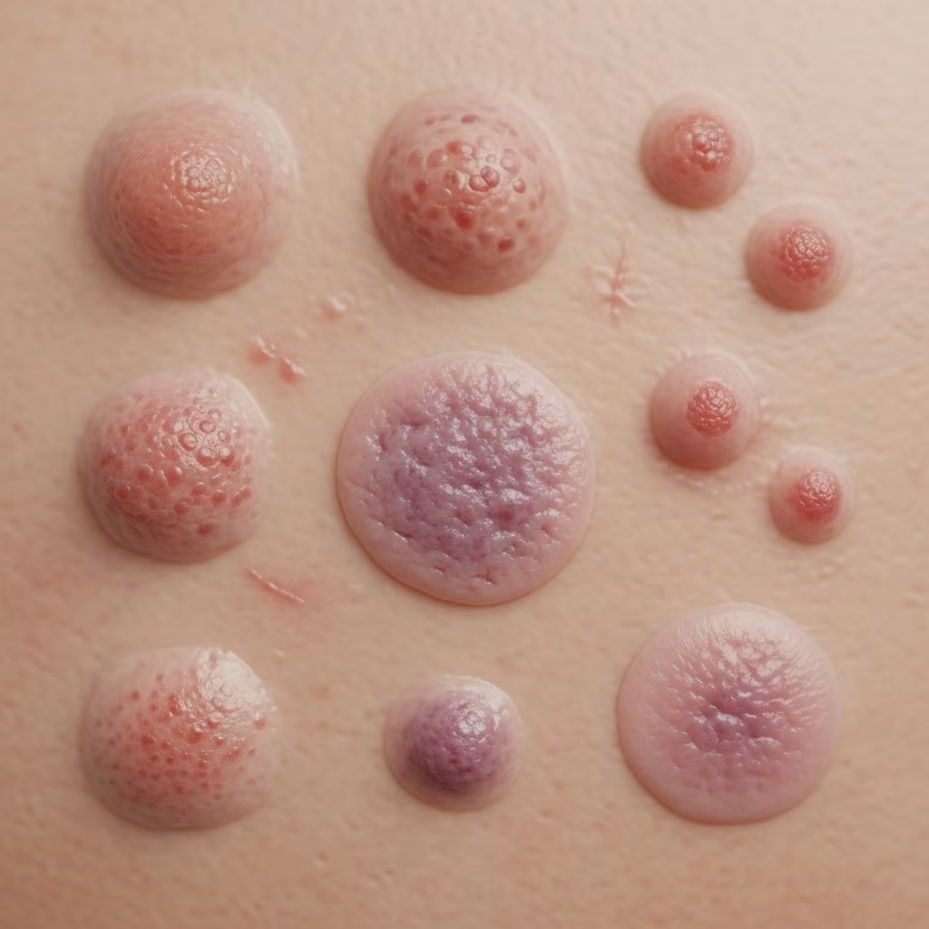
Sarcoidosis is a multisystem granulomatous disease with a wide array of clinical presentations, including notable skin manifestations. Early recognition and effective management of cutaneous sarcoidosis can aid in timely diagnosis, guide prognosis, and improve patients’ quality of life. This comprehensive article explores the clinical features, diagnostic strategies, differential diagnoses, and management of skin lesions in sarcoidosis using current evidence and clinical guidelines.
Introduction to Sarcoidosis and Skin Lesions
Sarcoidosis is a chronic, multisystem inflammatory disorder characterized by the formation of noncaseating granulomas in various tissues. Its etiology remains uncertain, but immune dysregulation and genetic predisposition are recognized factors. Of all potential sites, the skin is involved in approximately 20–35% of cases and often serves as a visible clue for systemic involvement. Cutaneous manifestations can be the first or only sign of disease, making their recognition crucial for diagnostic and prognostic evaluation.
Epidemiology and Significance of Cutaneous Involvement
Cutaneous involvement in sarcoidosis affects both sexes and a broad age range but is more prevalent in women and certain ethnic groups. In up to 30% of cases, skin lesions are the initial presenting feature. Recognition of skin lesions is critical not only because they hint at underlying systemic disease but also offer accessible sites for tissue diagnosis and potential prognostic indicators.
- Skin lesions may herald more severe systemic involvement, including respiratory or cardiac sarcoidosis.
- Some cutaneous variants are associated with a greater risk of chronic, progressive disease.
Types of Skin Lesions in Sarcoidosis
The skin manifestations of sarcoidosis are broadly divided into specific and nonspecific lesions, depending on histopathological findings:
| Type | Histological Feature | Clinical Common Examples |
|---|---|---|
| Specific lesions | Noncaseating granulomas | Papules, plaques, lupus pernio, nodules, infiltrative scars |
| Non-specific lesions | No granulomas | Erythema nodosum, calcinosis cutis, digital clubbing |
Specific lesions usually display noncaseating granulomas on skin biopsy, whereas nonspecific lesions do not.
Common Specific Lesions
- Papules: Small, raised bumps (often 1–5 mm), most frequently found on the face, especially around the eyes, nose, and mouth. Can be solitary or grouped.
- Plaques: Larger, flat-topped, indurated lesions that may be single or multiple, with distribution often on the face, trunk, or limbs. They can mimic chronic discoid lupus or psoriasis.
- Lupus pernio: Chronic, violaceous, infiltrated plaques and nodules, commonly affecting the nose, cheeks, lips, and ears, associated with chronic systemic disease.
- Subcutaneous nodules: Firm, painless nodules, often located on arms, legs, or trunk.
- Scar (infiltrative) sarcoidosis: Recurrence of sarcoidal granulomas within old scars or tattoos, typically as raised, violaceous areas.
- Uncommon morphologies: Ichthyosiform, atrophic, ulcerative, hypopigmented patches, alopecia, and mucosal involvement.
Common Non-Specific Lesions
- Erythema nodosum: Painful, red nodules typically on the anterior shins, often acute and self-limiting. Strongly associated with acute systemic sarcoidosis (Löfgren’s syndrome).
- Other rare presentations: Calcinosis cutis, prurigo, digital clubbing.
Clinical Recognition and Common Presentations
Cutaneous sarcoidosis presents a diagnostic challenge due to its diverse morphologies and ability to mimic other dermatologic diseases. Recognizing characteristic lesions is essential for early detection and management. The color of lesions may vary: reddish to violaceous, brown, translucent, or hyperpigmented. Induration is typical on palpation, especially for papular and plaque lesions. Some lesions may be disfiguring, especially those affecting the face or hands.
- Papules: May range from only a few lesions to multiple, widespread, grouped, or confluent papules. Commonly found on periorificial areas (around eyes, nostrils, mouth).
- Plaques: Often symmetric distribution when multiple, may present telangiectatic vessels, scaling, or mimic discoid lupus.
- Lupus pernio: Slowly progressive, chronic course; a strong predictor of chronic systemic disease, especially respiratory involvement.
- Erythema nodosum: Signals acute systemic sarcoidosis but not granulomatous infiltration of skin.
Diagnosis: Biopsy and Other Investigations
Gold-standard diagnosis of cutaneous sarcoidosis relies on histologic demonstration of noncaseating granulomas in the affected tissue, with exclusion of other granulomatous diseases. The diagnostic approach includes:
- Punch biopsy or incisional wedge biopsy of the skin lesion, incorporating sufficient dermal tissue for analysis.
- Histopathology: Presence of well-formed, uniform, noncaseating epithelioid cell granulomas, typically with scant surrounding lymphocytes (“naked tubercles”). Reticulin fibers may be seen around granulomas.
- Exclusion of infectious causes via culture or special stains (mycobacterial, fungal) when appropriate.
- Systemic assessment after skin confirmation: chest imaging, pulmonary function tests, serum ACE, calcium, and organ-specific workup as indicated.
The punch biopsy is highly effective, diagnostic in approximately 80% of cases, with a low complication rate. More extensive incisional or excisional biopsies may be considered if initial samples are nondiagnostic. A systemic evaluation is also critical to determine the full extent of organ involvement.
Differential Diagnosis of Skin Lesions
Due to their protean clinical presentations, cutaneous sarcoidosis lesions must be distinguished from a variety of other dermatological and systemic conditions. Histopathology, supported by clinical examination and history, is essential for accurate differentiation.
| Lesion Type | Lookalike Diseases |
|---|---|
| Papules | Granulomatous rosacea, acne, benign appendageal tumors |
| Plaques | Psoriasis, lichen planus, nummular eczema, discoid lupus erythematosus, granuloma annulare, T-cell lymphoma, Kaposi’s sarcoma, secondary syphilis |
| Lupus pernio | Sarcoidosis-specific; facial discoid lupus |
| Scar involvement | Hypertrophic scars, keloids |
| Erythema nodosum | Cellulitis, furunculosis, other causes of panniculitis |
On histology, key features—such as noncaseating granulomas with scant lymphocytes (as opposed to caseation or lymphocyte-heavy granulomas)—help distinguish sarcoidosis from leprosy, lupus vulgaris, and other granulomatous disorders.
Prognostic Implications of Specific Lesions
Recognition of the type of cutaneous lesion has significant prognostic value:
- Lupus pernio and chronic plaques frequently correlate with a more persistent and severe course, often indicating systemic involvement (especially respiratory system) and resistance to therapy.
- Erythema nodosum is associated with acute systemic forms (such as Löfgren’s syndrome), typically conferring a favorable prognosis with spontaneous resolution.
- High serum ACE levels, BAL lymphocytosis, and an increased CD4/CD8 lymphocyte ratio in patients with specific cutaneous lesions may signal a greater risk for systemic progression and worse outcomes.
Management Strategies for Skin Lesions
Cutaneous sarcoidosis treatment is tailored to the severity, extent, lesion type, systemic involvement, and impact on function or appearance. Lesions may spontaneously remit; thus, not all cases require intervention. Key management strategies include:
Local Treatment Options
- High-potency topical corticosteroids are the standard first-line therapy for limited disease; effective for papules, plaques, and small lesions.
- Intralesional corticosteroids (e.g., triamcinolone acetonide) are preferred for localized, nodular, or disfiguring lesions, such as lupus pernio.
- Topical calcineurin inhibitors (e.g., tacrolimus) as an alternative for those intolerant to steroids or with thinner skin involvement.
Systemic Treatment Options
- Oral corticosteroids (prednisone): Indicated for widespread or disfiguring skin involvement, or if accompanied by severe systemic disease. Tapering is gradual based on response.
- Steroid-sparing immunomodulators: Methotrexate, hydroxychloroquine, azathioprine, or mycophenolate mofetil, used for steroid-refractory or chronic cases.
- Anti-TNF-alpha agents (e.g., infliximab): Considered in select refractory cases, especially lupus pernio resistant to conventional therapies.
- Antimalarials (e.g., hydroxychloroquine): Also effective in cutaneous sarcoid, particularly when corticosteroids are contraindicated.
General Principles in Management
- Treat underlying systemic manifestations where applicable, using a multidisciplinary approach.
- Monitor for adverse effects and complications of immunosuppressive therapy.
- Patient education and regular follow-up are essential due to relapsing-remitting course.
Many patients experience spontaneous remission, but those with chronic or cosmetically significant lesions may require long-term management. The risk of skin atrophy with local corticoid use and systemic drug toxicity (hepatic, hematologic, ocular) must be weighed against benefits.
Psychosocial Impact of Cutaneous Sarcoidosis
While cutaneous sarcoidosis seldom results in serious morbidity or mortality, its visible and often disfiguring nature can lead to considerable psychosocial distress, anxiety, and diminished quality of life. Facial scarring or lesions, in particular, may cause embarrassment or stigmatization, highlighting the importance of sensitive management and patient support.
Frequently Asked Questions (FAQs)
Q: What are the most common cutaneous manifestations of sarcoidosis?
A: The most common skin lesions are papules, plaques, subcutaneous nodules, lupus pernio, and erythema nodosum. Papules and plaques are typical ‘specific’ granulomatous lesions, while erythema nodosum is a nonspecific reactive process often seen in acute presentations.
Q: How is cutaneous sarcoidosis diagnosed?
A: Diagnosis is made by a skin biopsy showing noncaseating granulomas, supported by clinical, radiologic, and laboratory evaluation to exclude other causes and assess systemic involvement.
Q: Are all skin lesions in sarcoidosis treated the same way?
A: No. Treatment is tailored according to severity, lesion morphology, location, patient preference, and associated systemic disease. Options range from topical/intralesional to systemic therapies.
Q: Can skin lesions in sarcoidosis resolve spontaneously?
A: Yes, especially non-disfiguring or nonspecific lesions such as erythema nodosum. Chronic or severe forms, such as lupus pernio, are less likely to resolve without treatment.
Q: What complications can occur from skin lesions?
A: Mainly cosmetic disfigurement and psychosocial stress. Rare complications include ulceration, secondary infection, or chronic lymphedema in severe plaque or lupus pernio cases.
References
- Clinical and Prognostic Significance of Skin Lesions in Sarcoidosis, PMC3614473
- Cutaneous Sarcoidosis: A Dermatologic Masquerader – AAFP
- Sarcoidosis and Its Dermatological Manifestations: A Narrative Review, PMC9481194
- Sarcoidosis and your skin: Diagnosis and treatment, American Academy of Dermatology
References
- https://pmc.ncbi.nlm.nih.gov/articles/PMC3614473/
- https://www.aafp.org/pubs/afp/issues/2002/0415/p1581.html
- https://pmc.ncbi.nlm.nih.gov/articles/PMC9481194/
- https://jamanetwork.com/journals/jamadermatology/fullarticle/2797292
- https://www.aad.org/public/diseases/a-z/sarcoidosis-treatment
- https://www.stopsarcoidosis.org/skin-sarcoidosis/
- https://www.mayoclinic.org/diseases-conditions/sarcoidosis/diagnosis-treatment/drc-20350363
- https://dermnetnz.org/topics/sarcoidosis