

Understanding Chronic Leg Ulcers
Chronic leg ulcers represent one of the most challenging wound care conditions, affecting millions of people worldwide and significantly impacting quality of life. These persistent wounds, primarily caused by venous insufficiency, arterial disease, or a combination of vascular problems, require specialized management approaches to achieve successful healing outcomes.
Venous leg ulcers constitute the majority of chronic leg ulcers, accounting for approximately 80% of all lower extremity wounds. These ulcers develop when the venous system fails to efficiently return blood from the legs back to the heart, leading to increased pressure in the veins and subsequent tissue damage. The resulting wounds are typically shallow, irregular in shape, and located around the medial malleolus area of the lower leg.
The complexity of chronic leg ulcers lies not only in their underlying pathophysiology but also in the multiple factors that can impede healing. These include bacterial colonization, excessive wound exudate, prolonged inflammation, and the presence of biofilms that protect bacteria from conventional treatments. Understanding these challenges is crucial for healthcare providers when selecting appropriate therapeutic interventions.
The Role of Therapeutic Dressings
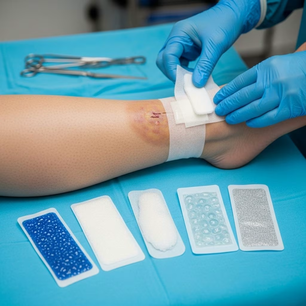
Therapeutic dressings serve as a cornerstone in the management of chronic leg ulcers, working in conjunction with compression therapy to create an optimal healing environment. Unlike traditional gauze dressings that merely cover wounds, modern therapeutic dressings actively participate in the healing process by addressing specific wound characteristics and patient needs.
The primary functions of therapeutic dressings include maintaining appropriate moisture levels in the wound bed, protecting against bacterial contamination, managing excessive exudate, promoting autolytic debridement of necrotic tissue, and providing thermal insulation to support cellular activity. Advanced dressings can also deliver therapeutic agents directly to the wound site, such as antimicrobial compounds or growth factors that accelerate healing.
Research has consistently demonstrated that maintaining a moist wound environment significantly accelerates healing compared to allowing wounds to dry out. Therapeutic dressings achieve this by creating a sealed environment that prevents excessive moisture loss while allowing for controlled vapor transmission. This balanced approach prevents both desiccation and maceration, two common complications that can delay healing in chronic wounds.
Types of Therapeutic Dressings
Hydrocolloid Dressings
Hydrocolloid dressings represent one of the most widely used therapeutic dressing categories for chronic leg ulcers. These dressings contain gel-forming agents such as pectin, gelatin, and carboxymethylcellulose that interact with wound exudate to form a gel-like substance over the wound bed. This gel maintains optimal moisture levels while providing a protective barrier against external contaminants.
The benefits of hydrocolloid dressings include their ability to promote autolytic debridement, reduce pain during dressing changes, and provide extended wear time of up to seven days in low-exuding wounds. They are particularly effective for chronic leg ulcers in the proliferative phase of healing when granulation tissue formation is the primary goal.
Foam Dressings
Foam dressings are highly absorbent polyurethane-based dressings designed to manage moderate to heavy wound exudate while maintaining a moist healing environment. Their unique structure allows for vertical wicking of fluid away from the wound bed while preventing lateral spread that could cause periwound skin maceration.
These dressings are ideal for venous leg ulcers that produce significant amounts of exudate, a common characteristic of these wounds. The cushioning effect of foam dressings also provides protection against external trauma and pressure, making them suitable for mobile patients who need to maintain their daily activities during treatment.
Alginate Dressings
Alginate dressings, derived from brown seaweed, possess exceptional absorptive capacity and hemostatic properties. When in contact with wound exudate, calcium alginate fibers transform into a soft gel that conforms to the wound bed, providing intimate contact with irregular wound surfaces common in chronic leg ulcers.
The high absorption capacity of alginate dressings makes them particularly suitable for heavily exuding wounds during the inflammatory phase of healing. Additionally, the calcium ions released during the gelling process can help control minor bleeding and may have beneficial effects on cellular activity involved in wound healing.
Hydrogel Dressings
Hydrogel dressings contain high water content (typically 80-90%) and are available in both sheet and amorphous forms. These dressings are designed to donate moisture to dry wound beds while providing cooling and soothing effects that can help reduce wound-related pain.
Hydrogel dressings are particularly valuable in the management of chronic leg ulcers with necrotic tissue or slough, as they facilitate autolytic debridement by softening and loosening devitalized tissue. The cooling effect of hydrogels can also provide significant pain relief, improving patient comfort and compliance with treatment regimens.
Vapor-Permeable Films and Membranes
These thin, transparent dressings allow for moisture vapor transmission while preventing bacterial penetration. Film dressings are primarily used as secondary dressings to secure primary dressings or to protect periwound skin from adhesive trauma.
While not typically used as primary dressings for chronic leg ulcers due to their limited absorptive capacity, film dressings play an important role in comprehensive wound management by providing a waterproof barrier that allows patients to shower while protecting the wound site.
Antimicrobial Dressings and Topical Agents
Silver-Based Dressings
Silver-containing dressings have gained significant popularity in chronic wound management due to their broad-spectrum antimicrobial properties. These dressings release silver ions in a controlled manner, providing sustained antimicrobial activity against bacteria, fungi, and some viruses that commonly colonize chronic wounds.
Various forms of silver dressings are available, including silver sulfadiazine cream, nanocrystalline silver dressings, and silver-impregnated foam or alginate dressings. Clinical studies have shown that silver dressings can effectively reduce bacterial load in chronic wounds, though their impact on healing rates requires further investigation.
Iodine-Containing Products
Iodine-based antimicrobial agents, particularly cadexomer iodine and povidone-iodine, have demonstrated effectiveness in managing infected chronic leg ulcers. Cadexomer iodine combines the antimicrobial properties of iodine with the absorptive capacity of cadexomer beads, making it particularly suitable for infected, exuding wounds.
Research has shown that iodine-containing dressings can effectively reduce methicillin-resistant Staphylococcus aureus (MRSA) and Pseudomonas aeruginosa, two problematic organisms commonly found in chronic wounds. The broad-spectrum antimicrobial activity of iodine, combined with its low toxicity profile, makes it an attractive option for long-term wound management.
Chlorhexidine-Impregnated Dressings
Chlorhexidine is another antimicrobial agent that has been incorporated into wound dressings for managing colonized or infected chronic leg ulcers. While chlorhexidine demonstrates good antimicrobial activity and has been associated with few adverse effects on wound healing, current evidence regarding its efficacy in chronic wound management remains limited.
Healthcare providers should carefully consider the risk-benefit profile when selecting chlorhexidine-containing dressings, as more robust clinical data is needed to establish clear recommendations for their use in chronic leg ulcer management.
Dressing Selection Criteria
Selecting the most appropriate therapeutic dressing for chronic leg ulcers requires careful assessment of multiple wound and patient factors. The decision-making process should be systematic and evidence-based to optimize healing outcomes while considering cost-effectiveness and patient preferences.
Wound Assessment Parameters
The first step in dressing selection involves comprehensive wound assessment, including measurement of wound dimensions, evaluation of wound bed characteristics, assessment of exudate levels, identification of signs of infection, and examination of periwound skin condition. Each of these parameters influences dressing choice and treatment strategy.
Wound bed assessment should focus on the percentage of granulation tissue, presence of slough or necrotic tissue, and overall wound appearance. Heavily necrotic wounds may benefit from debriding agents or dressings that promote autolytic debridement, while clean, granulating wounds require dressings that support tissue proliferation and epithelialization.
Exudate Management
Exudate levels significantly influence dressing selection, as inappropriate management can lead to either wound desiccation or periwound maceration. Low-exuding wounds typically benefit from hydrocolloid or hydrogel dressings that maintain moisture, while heavily exuding wounds require highly absorbent dressings such as foams or alginates.
The composition of wound exudate also provides valuable information about wound status. Purulent or malodorous exudate may indicate infection, warranting the use of antimicrobial dressings in conjunction with appropriate systemic therapy.
| Exudate Level | Recommended Dressing Types | Key Considerations |
|---|---|---|
| Minimal | Hydrocolloid, Film dressings | Maintain moisture, prevent desiccation |
| Low | Hydrocolloid, Hydrogel | Support autolytic debridement |
| Moderate | Foam, Hydrocolloid with border | Balance absorption with moisture retention |
| Heavy | Alginate, High-absorbent foam | Prevent periwound maceration |
| Very Heavy | Super-absorbent dressings, NPWT | Consider underlying pathology |
Comprehensive Treatment Approach
Effective management of chronic leg ulcers extends beyond dressing selection to encompass a holistic treatment approach that addresses underlying pathophysiology, patient factors, and lifestyle modifications. Therapeutic dressings work most effectively when integrated into a comprehensive care plan.
Compression Therapy Integration
Compression therapy remains the gold standard treatment for venous leg ulcers and should be used in conjunction with appropriate wound dressings. The selection of dressing type may influence the compression system used, as some dressings require more frequent changes that could interfere with compression bandage integrity.
Healthcare providers must ensure that chosen dressings are compatible with compression systems and that the combined approach does not compromise either treatment modality. Multi-layer compression systems may accommodate bulkier dressings better than single-layer systems.
Wound Debridement Strategies
Debridement of necrotic tissue and debris is often necessary to create an optimal healing environment for chronic leg ulcers. The choice of debridement method can influence dressing selection, as some dressings actively participate in the debridement process.
Autolytic debridement using moisture-retentive dressings such as hydrocolloids or hydrogels offers a gentle, pain-free approach suitable for patients who cannot tolerate more aggressive debridement methods. Enzymatic debridement using collagenase-containing ointments can be combined with appropriate cover dressings to enhance efficacy.
Application Techniques and Best Practices
Proper application technique is crucial for maximizing the therapeutic benefits of wound dressings while minimizing complications such as periwound skin damage or premature dressing failure. Healthcare providers should follow manufacturer guidelines while adapting techniques to individual patient needs.
Preparation and Cleansing
Before dressing application, the wound and surrounding skin should be gently cleansed with appropriate solutions to remove debris and excess exudate. Normal saline remains the gold standard for wound irrigation, though other solutions may be appropriate in specific circumstances.
Periwound skin should be assessed for signs of maceration, dermatitis, or allergic reactions that might influence dressing choice or require additional protective measures. Barrier products may be necessary to protect vulnerable periwound skin from adhesive trauma or excessive moisture exposure.
Sizing and Securing
Dressing size should extend approximately 2-3 centimeters beyond the wound edges to ensure adequate coverage while avoiding excessive overlap onto healthy skin. Oversized dressings can cause unnecessary adhesive trauma, while undersized dressings may allow wound edges to dry out or become contaminated.
Secondary dressings or retention devices should be used as needed to secure primary dressings without compromising circulation or causing pressure points. The choice of securing method should consider patient activity level, anatomical location, and skin integrity.
Monitoring and Assessment
Regular monitoring and assessment of wound progress is essential for optimizing therapeutic outcomes and identifying the need for treatment modifications. Healthcare providers should establish systematic evaluation protocols that include both objective measurements and subjective patient reporting.
Healing Progress Indicators
Key indicators of healing progress include reduction in wound dimensions, improvement in wound bed appearance, decreased exudate levels, and absence of clinical signs of infection. Digital photography can be valuable for documenting changes over time and facilitating communication between healthcare team members.
Lack of healing progress after 4-6 weeks of appropriate treatment should prompt reassessment of the diagnosis, treatment plan, and patient adherence. Consideration should be given to underlying factors that may be impeding healing, such as inadequate compression, undiagnosed arterial disease, or nutritional deficiencies.
Complication Recognition
Healthcare providers must be vigilant for signs of complications that may arise during treatment, including contact dermatitis from dressing materials, periwound skin maceration from excessive moisture, and signs of wound infection that may require systemic intervention.
Patient education regarding warning signs and when to seek medical attention is crucial for preventing serious complications and ensuring timely intervention when problems arise.
Frequently Asked Questions (FAQs)
Q: How often should therapeutic dressings be changed on chronic leg ulcers?
A: Dressing change frequency depends on the type of dressing used and wound characteristics. Hydrocolloid dressings may be left in place for up to 7 days if there are no signs of infection or excessive exudate. Foam dressings typically require changing every 3-5 days, while alginate dressings may need daily changes in heavily exuding wounds. Always follow manufacturer guidelines and clinical assessment.
Q: Can I shower or bathe with therapeutic dressings in place?
A: Some therapeutic dressings are waterproof and allow for showering, while others may require protection with a waterproof cover or removal before bathing. Film dressings and some foam dressings with adhesive borders can typically withstand brief water exposure. Check with your healthcare provider about the specific dressing being used and follow their recommendations.
Q: What should I do if the skin around my leg ulcer becomes red or irritated?
A: Periwound skin irritation may indicate an allergic reaction to the dressing, excessive moisture exposure, or incorrect application technique. Contact your healthcare provider immediately if you notice increasing redness, swelling, warmth, or pain around the wound. They may need to change the dressing type or provide additional skin protection measures.
Q: Are antimicrobial dressings always necessary for chronic leg ulcers?
A: Antimicrobial dressings are not routinely required for all chronic leg ulcers. They are typically reserved for wounds showing signs of infection, critical colonization, or delayed healing despite appropriate standard care. Overuse of antimicrobial dressings can lead to resistance and may not be cost-effective. Your healthcare provider will determine if antimicrobial therapy is appropriate for your specific situation.
Q: How long does it typically take for a chronic leg ulcer to heal with proper dressing management?
A: Healing time varies significantly depending on wound size, depth, patient factors, and adherence to treatment. Small ulcers may heal within 6-12 weeks with appropriate care, while larger or more complex wounds may take several months. Factors such as adequate compression therapy, nutrition, blood sugar control, and smoking cessation significantly impact healing rates. Regular follow-up with healthcare providers is essential for monitoring progress.
References
- https://pmc.ncbi.nlm.nih.gov/articles/PMC4144245/
- https://pmc.ncbi.nlm.nih.gov/articles/PMC6513558/
- https://www.asgfoot.com/post/early-treatment-venous-leg-ulcers-effective-healing
- https://www.nhs.uk/conditions/leg-ulcer/treatment/
- https://legsmatter.org/information-and-support/treatment-options/wound-dressings/
- https://www.aafp.org/pubs/afp/issues/2010/0415/p989.html
- https://www.racgp.org.au/afp/2014/september/ulcer-dressings-and-management
- https://absorbest.com/leg-ulcer-treatment/