

Table of Contents
\n
- \n
- Understanding Acne Lesions
- Papules: The Early Inflammatory Stage
- Pustules: When Infection Takes Hold
- Nodules: Deep Cystic Formations
- Acne Severity Grading Systems
- Treatment Approaches by Lesion Type
- Prevention and Management Strategies
- When to See a Dermatologist
- Frequently Asked Questions
\n
\n
\n
\n
\n
\n
\n
\n
\n
\n
\n\n
Acne affects millions of people worldwide, creating not just physical discomfort but often emotional distress as well. Understanding the different types of acne lesions is crucial for effective treatment and prevention of scarring. While many people use the term \”pimple\” generically, dermatologists classify acne lesions into specific categories based on their appearance, depth, and inflammatory characteristics. This comprehensive guide explores the key differences between papules, pustules, and nodules, helping you identify what type of acne you’re dealing with and how to treat it appropriately.
\n\n
Understanding Acne Lesions
\n\n
Acne vulgaris is a complex skin condition that develops when hair follicles become clogged with oil, dead skin cells, and bacteria. The process begins deep within the pilosebaceous unit, where sebaceous glands produce sebum to lubricate the skin. When this natural process goes awry, various types of lesions can form, each representing a different stage or severity of the inflammatory process.
\n\n
Acne lesions are broadly categorized into two main types: non-inflammatory and inflammatory. Non-inflammatory lesions include comedones (blackheads and whiteheads), while inflammatory lesions encompass papules, pustules, nodules, and cysts. The transition from non-inflammatory to inflammatory acne occurs when the follicle wall ruptures, releasing its contents into the surrounding tissue and triggering an immune response.
\n\n
The Formation Process
\n\n
The development of acne lesions follows a predictable pattern. Initially, excess sebum production combined with abnormal keratinization leads to the formation of a microcomedo. As this plug grows and becomes exposed to air, it may develop into an open comedone (blackhead). If the follicle remains closed, a closed comedone (whitehead) forms. When bacteria, particularly Propionibacterium acnes, proliferate within the blocked follicle, inflammation ensues, leading to the formation of papules, pustules, and in severe cases, nodules and cysts.
\n\n
Papules: The Early Inflammatory Stage
\n\n
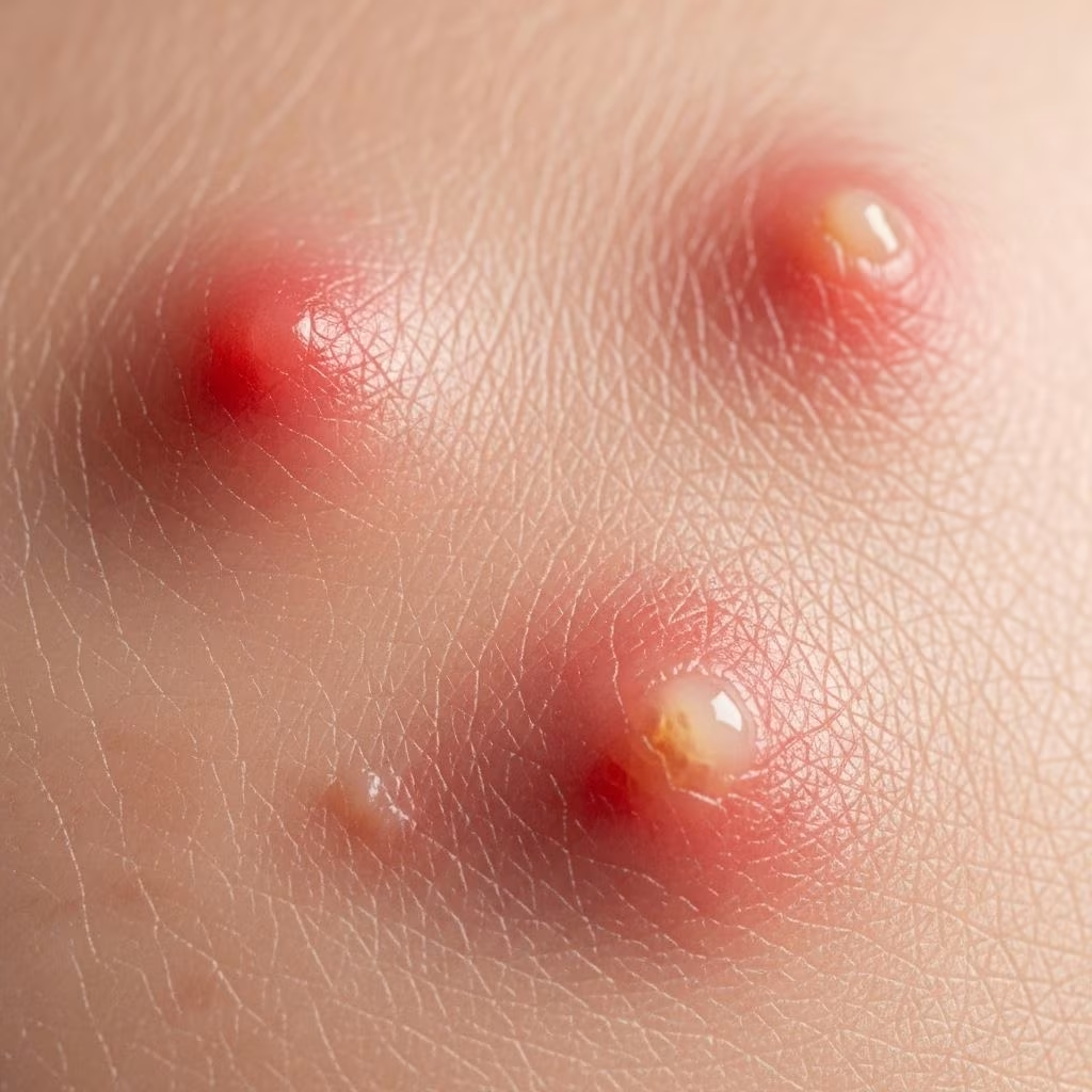
Papules represent the earliest stage of inflammatory acne and are characterized by small, raised bumps on the skin that are typically red or pink in color. These lesions measure less than 5 millimeters in diameter and do not contain visible pus, distinguishing them from pustules. Papules form when the follicle wall breaks down due to the pressure from accumulated sebum and keratin, allowing bacteria and inflammatory substances to escape into the surrounding dermis.
\n\n
Physical Characteristics of Papules
\n\n
Papules appear as firm, dome-shaped elevations on the skin surface. They are typically tender to touch and may feel warm due to the underlying inflammation. The surrounding skin often shows erythema (redness), which can extend beyond the actual lesion boundaries. Unlike pustules, papules lack a visible white or yellow center, as the inflammatory process has not yet produced enough purulent material to become visible on the surface.
\n\n
The color of papules can vary depending on skin tone and the degree of inflammation. In lighter skin types, they typically appear bright red, while in darker skin tones, they may appear as hyperpigmented bumps or maintain a reddish hue. The inflammation associated with papules can persist for several days to weeks if left untreated.
\n\n
Location and Distribution
\n\n
Papules commonly occur in areas with high concentrations of sebaceous glands, including the face (particularly the T-zone), chest, upper back, and shoulders. They may appear as isolated lesions or in clusters, depending on the severity of the acne condition. The distribution pattern often reflects hormonal influences, with adolescents typically experiencing more facial involvement, while adults may see more lesions on the lower face, jawline, and neck.
\n\n
Pustules: When Infection Takes Hold
\n\n
Pustules develop when papules progress further in the inflammatory process, accumulating enough purulent material to become visible on the skin surface. These lesions are characterized by a raised, inflamed base topped with a white or yellow center containing pus. Pustules represent a more advanced stage of inflammatory acne and indicate increased bacterial activity within the follicle.
\n\n
Formation and Development
\n\n
The transition from papule to pustule occurs when neutrophils (white blood cells) migrate to the site of inflammation in response to bacterial overgrowth and toxic substances produced by P. acnes. These immune cells attempt to combat the infection by releasing enzymes and producing pus, a mixture of dead bacteria, neutrophils, and cellular debris. The accumulation of this purulent material creates the characteristic white or yellow head that distinguishes pustules from papules.
\n\n
Pustules typically measure between 5 to 10 millimeters in diameter, though they can sometimes be larger. The inflammatory base extends deeper into the dermis compared to papules, making pustules more prone to scarring if manipulated or improperly treated. The pressure from accumulated pus can cause significant discomfort and may lead to spontaneous rupture if the lesion becomes too large.
\n\n
Clinical Appearance and Symptoms
\n\n
Pustules present as elevated lesions with a distinct inflammatory component. The base is typically red, swollen, and tender to touch, while the central portion contains visible pus that may range from white to yellow or even greenish in color. The surrounding skin often shows significant erythema and may feel warm due to increased blood flow to the area.
\n\n
Patients often experience more discomfort with pustules compared to papules, including pain, throbbing, and increased sensitivity. The temptation to squeeze or pop pustules is common but strongly discouraged, as this can spread bacteria to surrounding tissue, worsen inflammation, and increase the risk of scarring.
\n\n
Nodules: Deep Cystic Formations
\n\n
Nodules represent the most severe form of inflammatory acne and are characterized by large, deep, hard lumps that develop beneath the skin surface. These lesions extend well into the dermis and subcutaneous tissue, measuring greater than 10 millimeters in diameter. Nodules form when the inflammatory process spreads extensively through the dermis, creating large areas of tissue destruction and intense immune response.
\n\n
Pathophysiology of Nodular Acne
\n\n
The development of nodular acne involves complete rupture of the follicle wall deep within the dermis, allowing sebum, keratin, and bacteria to spread extensively through surrounding tissue. This triggers a massive inflammatory response involving multiple cell types, including macrophages, lymphocytes, and giant cells. The body attempts to wall off the infection by forming a fibrous capsule around the affected area, creating the characteristic hard, immovable nodule.
\n\n
Nodular acne often develops from pre-existing inflammatory lesions that have been inadequately treated or manipulated. The deep location and extensive inflammatory component make nodules particularly resistant to topical treatments and more likely to cause permanent scarring. The healing process is prolonged, often taking weeks to months for complete resolution.
\n\n
Clinical Features and Complications
\n\n
Nodules appear as large, firm, often painful lumps that may or may not have a visible surface component. Unlike pustules, nodules rarely come to a head and cannot be easily expressed. They may feel like hard marbles under the skin and can cause significant disfigurement, particularly when multiple nodules occur in the same area.
\n\n
The pain associated with nodular acne can be severe and may interfere with daily activities, especially when nodules occur in areas that are frequently touched or rubbed by clothing. The deep inflammatory process often results in post-inflammatory hyperpigmentation that can persist for months after the lesion has healed. Nodular acne carries the highest risk of scarring among all acne types and may result in ice pick scars, rolling scars, or hypertrophic scarring.
\n\n
Acne Severity Grading Systems
\n\n
Dermatologists use various grading systems to assess acne severity and guide treatment decisions. Understanding these classifications helps both patients and healthcare providers communicate effectively about treatment goals and expectations. The most commonly used systems consider the type, number, and distribution of lesions.
\n\n
Standard Severity Classifications
\n\n
| Severity Level | Comedones | Inflammatory Lesions | Total Lesions | Special Features |
|---|---|---|---|---|
| Mild | < 20 | < 15 | < 30 | Scattered lesions, minimal scarring |
| Moderate | 20-100 | 15-50 | 30-125 | More widespread, some scarring |
| Severe | > 100 | > 50 | > 125 | > 5 cysts, significant scarring risk |
\n\n
Lesion-Specific Grading
\n\n
Beyond numerical counts, dermatologists also consider the specific types of lesions present when determining severity. The presence of nodules automatically classifies acne as severe, regardless of the total lesion count. Similarly, any acne that causes scarring or significant psychological distress may warrant more aggressive treatment typically reserved for severe cases.
\n\n
The Global Acne Grading System incorporates both lesion count and anatomical distribution, recognizing that acne affecting larger surface areas or multiple body regions may require more intensive treatment. This system assigns point values to different body areas and lesion types, providing a comprehensive severity score.
\n\n
Treatment Approaches by Lesion Type
\n\n
Effective acne treatment requires matching the therapeutic approach to the specific type and severity of lesions present. While some treatments are effective across multiple lesion types, others are specifically targeted toward particular stages of the acne development process.
\n\n
Papule Treatment Strategies
\n\n
Papular acne typically responds well to topical treatments that address inflammation and bacterial overgrowth. Topical retinoids are considered first-line therapy as they help normalize follicular keratinization and reduce inflammation. Benzoyl peroxide provides antibacterial action and can be used alone or in combination with topical antibiotics like clindamycin.
\n\n
Anti-inflammatory treatments such as topical corticosteroids may be used for short-term management of severe inflammatory papules, though long-term use should be avoided due to potential side effects. Salicylic acid-based products can help reduce inflammation and promote gentle exfoliation of the skin surface.
\n\n
Pustule Management
\n\n
Pustular acne requires more aggressive antibacterial treatment due to the increased bacterial load. Combination therapies using benzoyl peroxide with topical antibiotics are highly effective. Topical retinoids remain important for preventing new lesion formation and should be continued even after active pustules have resolved.
\n\n
For moderate to severe pustular acne, oral antibiotics such as tetracyclines (doxycycline, minocycline) may be necessary. These medications provide both antibacterial and anti-inflammatory effects. Treatment duration typically ranges from 3-6 months, with careful monitoring for potential side effects and antibiotic resistance.
\n\n
Nodular Acne Treatment
\n\n
Nodular acne represents a treatment challenge and often requires systemic therapy. Oral isotretinoin is considered the gold standard treatment for severe nodular acne, providing comprehensive action against all factors involved in acne formation. However, due to its potential serious side effects, isotretinoin requires careful monitoring and is typically reserved for cases that have failed other treatments.
\n\n
Intralesional corticosteroid injections can provide rapid improvement for individual large nodules, reducing inflammation and preventing scarring. This treatment is performed in a dermatologist’s office and may need to be repeated for optimal results.
\n\n
Prevention and Management Strategies
\n\n
Preventing the progression of acne lesions from one type to another is often more effective than treating established lesions. A comprehensive prevention strategy addresses the multiple factors that contribute to acne development and helps maintain long-term skin health.
\n\n
Daily Skincare Routine
\n\n
Establishing a consistent skincare routine appropriate for acne-prone skin is fundamental to prevention. This includes gentle cleansing with non-comedogenic products, appropriate moisturization to maintain skin barrier function, and consistent use of topical treatments as prescribed. Over-washing or using harsh scrubbing can worsen inflammation and should be avoided.
\n\n
Sun protection is crucial for all acne patients, particularly those using photosensitizing medications like retinoids or tetracyclines. Non-comedogenic, broad-spectrum sunscreens should be used daily to prevent post-inflammatory hyperpigmentation and reduce the risk of scarring.
\n\n
Lifestyle Modifications
\n\n
Certain lifestyle factors can influence acne severity and lesion development. Stress management is important, as stress hormones can worsen acne. Regular exercise can help reduce stress and improve overall health, though proper hygiene after sweating is essential to prevent pore blockage.
\n\n
Dietary considerations may play a role in some individuals, with high-glycemic foods and dairy products potentially worsening acne in susceptible people. However, dietary modifications should complement, not replace, appropriate medical treatment.
\n\n
When to See a Dermatologist
\n\n
While mild acne can often be managed with over-the-counter treatments, certain situations warrant professional dermatological care. Understanding when to seek expert help can prevent progression to more severe lesions and reduce the risk of permanent scarring.
\n\n
Red Flags Requiring Professional Care
\n\n
The presence of nodular lesions always requires dermatological evaluation and treatment. Similarly, any acne that is causing scarring or significant psychological distress should be professionally managed, regardless of apparent severity. Acne that fails to respond to appropriate over-the-counter treatment after 6-8 weeks also warrants professional assessment.
\n\n
Signs of secondary bacterial infection, such as increased pain, warmth, red streaking, or systemic symptoms like fever, require immediate medical attention. These complications can occur when acne lesions become secondarily infected with other bacteria beyond the typical P. acnes.
\n\n
Frequently Asked Questions
\n\n
Q: Can I pop a pustule safely at home?
\n
A: It’s strongly advised against popping pustules at home as this can spread bacteria, worsen inflammation, and increase scarring risk. If extraction is necessary, it should be performed by a trained professional using sterile techniques.
\n\n
Q: How long do nodules take to heal?
\n
A: Nodules can take several weeks to months to fully resolve, even with appropriate treatment. The deep inflammatory nature of these lesions means healing is typically much slower than with superficial papules or pustules.
\n\n
Q: Are papules less serious than pustules?
\n
A: While papules represent an earlier stage of inflammation, both require appropriate treatment to prevent progression. Papules can develop into pustules if bacterial overgrowth occurs, so early intervention is important.
\n\n
Q: Can nodular acne be prevented?
\n
A: Early and appropriate treatment of mild to moderate acne can help prevent progression to nodular lesions. Consistent use of prescribed treatments and avoiding manipulation of existing lesions are key prevention strategies.
\n\n
Q: Do all types of acne leave scars?
\n
A: Not all acne lesions cause permanent scarring. However, the risk increases with lesion depth and severity. Nodular acne carries the highest scarring risk, while properly treated papules and pustules are less likely to cause permanent marks.
\n\n
Q: How can I tell the difference between a papule and a pustule?
\n
A: The main difference is the presence of visible pus. Papules are solid, raised bumps without a white or yellow center, while pustules have a visible pus-filled head. Both are inflamed and tender, but pustules indicate more advanced bacterial activity.
References
- https://www.aafp.org/pubs/afp/issues/2019/1015/p475.html
- https://www.merckmanuals.com/professional/multimedia/table/classification-of-acne-severity
- https://www.msdmanuals.com/professional/multimedia/table/classification-of-acne-severity
- https://apaxmedical.com/acne-grading-scale/
- https://www.ncbi.nlm.nih.gov/books/NBK459173/
- https://dermnetnz.org/topics/acne-vulgaris
- https://www.spiedigitallibrary.org/conference-proceedings-of-spie/9785/1/Acne-image-analysis-lesion-localization-and-classification/10.1117/12.2216444.full
- https://opendermatologyjournal.com/VOLUME/14/PAGE/38/
- https://www.nature.com/articles/s41598-024-84670-z