

Neurons, or nerve cells, are the building blocks of the nervous system. They are highly specialized cells responsible for receiving, processing, and transmitting information through electrochemical signals. Understanding the detailed structure of a neuron reveals how our brains and bodies communicate, control functions, and sense the world.
What Is a Neuron?
A neuron is a specialized cell in the nervous system designed to transmit information by both electrical and chemical means. Unlike most other cells in the body, neurons have unique extensions that enable them to connect and communicate over long distances. Their functional design allows the rapid and precise relay of data needed for thought, movement, sensation, and countless other bodily processes.
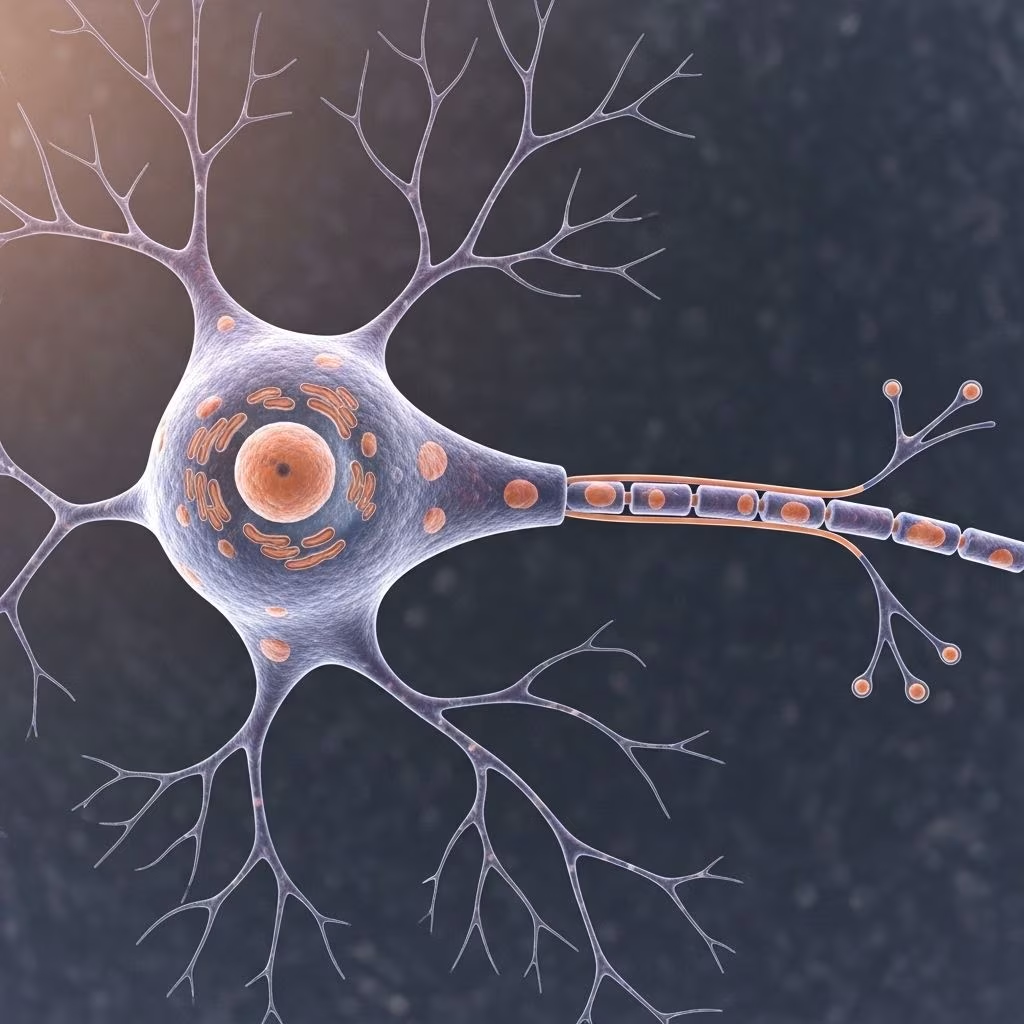
Every neuron, despite differences in type and function, shares a similar basic structure comprised of a cell body (soma), dendrites, and a single axon .
Core Parts of a Neuron
The anatomy of a neuron can be divided into several fundamental regions, each with distinct roles:
- Cell Body (Soma): Contains the nucleus and much of the cytoplasm; the metabolic center of the cell.
- Dendrites: Branching extensions that receive signals from other neurons.
- Axon: A long, thin fiber responsible for transmitting signals away from the cell body to other neurons, muscles, or glands.
- Axon Terminals: The endpoints of the axon, where communication with other cells occurs.
- Myelin Sheath: An insulating layer that covers some axons to enhance signal transmission speed.
- Nodes of Ranvier: Regularly spaced gaps in the myelin sheath critical for rapid signal conduction.
- Synapses: Junctions where the neuron communicates with another cell via neurotransmitters.
Detailed Anatomy of a Neuron
Cell Body (Soma)
The cell body, or soma, is the core part of the neuron. It contains the nucleus, where the neuron’s genetic material is stored, as well as essential organelles such as mitochondria (energy production), Golgi apparatus (protein processing), and endoplasmic reticulum (protein and lipid synthesis) .
- Function: Maintains overall neuron health, provides energy, synthesizes proteins, and integrates incoming signals.
- Anatomical Features: Surrounded by a membrane for protection and environmental interaction. Typical soma diameter ranges from 4 to 100 micrometers .
Dendrites
Dendrites are tree-like structures branching from the cell body. Their main role is to receive incoming signals (electrochemical messages) from other neurons or sensory receptors and transmit them toward the cell body .
- Structure: Can be highly branched, forming what is known as a dendritic tree.
- Function: Increase the surface area for receiving signals; covered with synapses for input from neighboring cells.
- Diversity: Some neurons, such as Purkinje cells in the cerebellum, have extremely elaborate dendritic trees capable of receiving thousands of signals at once .
Axon
The axon is a single elongated projection from the cell body that can extend up to a meter in length in humans. Its primary role is to convey electrical impulses, known as action potentials, away from the cell body toward other neurons or target tissues .
- Structure: Typically only one axon per neuron; branches might exist at the terminal end.
- Specialized Area: The axon hillock is where the axon joins the cell body—a critical site for action potential initiation due to a high density of ion channels .
- Axon Terminals: Small branches at the end of the axon that form junctions (synapses) with other neurons or effector cells.
Myelin Sheath and Nodes of Ranvier
The myelin sheath is a fatty, insulating layer surrounding the axon in many types of neurons. It is formed by specialized glial cells—Schwann cells in the peripheral nervous system and oligodendrocytes in the central nervous system .
- Function: Increases the speed and efficiency of electrical signal (action potential) transmission along the axon.
- Nodes of Ranvier: Small gaps between each segment of myelin sheath. These allow the nerve impulse to “jump” rapidly from node to node, a process called saltatory conduction, greatly enhancing transmission speed .
Synapse
A synapse is a complex junction where the axon terminal of one neuron comes in close contact with another cell—either another neuron, muscle cell, or gland cell. Here, information is transmitted chemically (via neurotransmitters) or, less commonly, electrically .
- Components: The presynaptic terminal (sending side), the synaptic cleft (the gap), and the postsynaptic membrane (receiving side).
- Function: When an action potential reaches the axon terminal, it causes the release of neurotransmitter molecules which cross the synaptic cleft and bind to receptors on the postsynaptic cell, initiating a new signal.
Table: Major Parts of a Neuron and Their Functions
| Part | Structure/Location | Main Function |
|---|---|---|
| Cell Body (Soma) | Contains nucleus; at core of neuron | Maintains cell function; integrates signals |
| Dendrites | Branch-like projections from soma | Receive incoming signals |
| Axon | Long, thin projection from soma | Transmits electrical impulses away from soma |
| Myelin Sheath | Fatty insulation along axon | Increases conduction speed |
| Nodes of Ranvier | Gaps in myelin sheath along axon | Allow rapid impulse transmission |
| Axon Terminals | Ends of axon branches | Release neurotransmitters; form synapses |
| Synapse | Junction with another cell | Information transfer between cells |
How Neurons Communicate
Electrical and Chemical Signals
Neuronal communication relies on two major processes:
- Electrical signaling: The movement of ions across the neuron’s membrane leads to a change in electrical charge, generating action potentials that travel along the axon.
- Chemical signaling: At the synapse, action potentials trigger the release of chemical messengers called neurotransmitters that bind to receptors on the target cell, transmitting or modulating the signal.
The Action Potential
A key feature of neurons is their ability to generate and propagate an action potential—a brief, rapid electrical impulse that travels down the axon. This is initiated at the axon hillock when signals received by dendrites and the cell body reach a threshold intensity .
- The action potential is all-or-none: it either occurs fully or not at all.
- As it travels along the axon, the action potential triggers the opening and closing of ion channels.
Synaptic Transmission
At the axon terminal, arrival of the action potential causes neurotransmitter-filled vesicles to merge with the presynaptic membrane and release their contents. Neurotransmitters cross the synaptic cleft, bind to receptors on the postsynaptic cell, and either excite or inhibit the new cell, depending on the type of neurotransmitter and receptor involved .
- Synapses can connect neurons to neurons, neurons to muscles (neuromuscular junctions), or neurons to glands (neuroeffector junctions).
- Each neuron may form thousands of synaptic connections, contributing to the vast complexity of the nervous system.
Types of Neurons
While all neurons share the same basic design, they can be classified based on structure, function, and location:
- Sensory (Afferent) Neurons: Transmit sensory information from receptors (e.g., skin, eyes) toward the central nervous system.
- Motor (Efferent) Neurons: Send commands from the central nervous system to muscles or glands.
- Interneurons: Connect neurons within the brain and spinal cord and play integrative roles.
Supporting Cells: Glial Cells
Neurons do not function in isolation; support is provided by glial cells:
- Schwann Cells: Produce the myelin sheath in the peripheral nervous system.
- Oligodendrocytes: Produce the myelin sheath in the central nervous system.
- Other glia: Astrocytes, microglia, and ependymal cells offer metabolic, structural, and immunological support.
Frequently Asked Questions (FAQs)
What are the main parts of a neuron, and what do they do?
A neuron’s main parts are the cell body (maintains cell health), dendrites (receive signals), and axon (transmits signals to other cells). The myelin sheath insulates the axon to speed signals, and synapses allow neurons to communicate chemically.
Why is the myelin sheath important?
The myelin sheath acts as insulation, allowing electrical signals to travel more quickly and efficiently along the axon. Without myelin, signals would travel more slowly, which could result in impaired movement, sensation, or cognition.
How do neurons communicate with each other?
Neurons communicate through synapses, where electrical impulses arriving at the axon terminal trigger the release of neurotransmitters. These chemicals cross to the next cell, where they generate a new electrical impulse or modify cell behavior.
Can a neuron have more than one axon?
Generally, neurons have only one axon, although the axon can branch extensively to communicate with many target cells. Multiple axons from a single neuron are rare.
What happens if parts of a neuron are damaged?
Damage to any part of a neuron can disrupt neural communication. For example, damage to the axon can block signal transmission, while loss of myelin (as in multiple sclerosis) slows or stops nerve impulses. Damage to dendrites or the cell body can impair the neuron’s ability to receive or integrate signals.
Key Takeaways
- Neurons are specialized nerve cells that process and transmit signals in the nervous system.
- Three core components—the cell body, dendrites, and axon—enable information flow and processing.
- Myelin sheath and nodes of Ranvier are critical to efficient signal transmission.
- Synapses are specialized junctions for communication between neurons and other cells via neurotransmitters.
- Supporting glial cells provide insulation, maintenance, and protection for neurons.
Further Reading
- Explore more about neuroanatomy and different neuron types to deepen your understanding of how the brain and nervous system work.
- Investigate neurological diseases, such as multiple sclerosis, to learn how disruptions in neuron structure and function affect the nervous system.
References
- https://bio.libretexts.org/Bookshelves/Human_Biology/Human_Biology_(Wakim_and_Grewal)/11:_Nervous_System/11.3:_Neurons
- https://www.healthline.com/health/neurons
- https://www.simplypsychology.org/neuron.html
- https://en.wikipedia.org/wiki/Neuron
- https://www.khanacademy.org/science/biology/human-biology/neuron-nervous-system/a/overview-of-neuron-structure-and-function
- https://byjus.com/biology/diagram-of-neuron/
- https://qbi.uq.edu.au/brain/brain-anatomy/types-neurons
- https://www.youtube.com/watch?v=6qS83wD29PY
- https://nba.uth.tmc.edu/neuroscience/m/s1/chapter08.html