

Conjunctivochalasis is a prevalent yet frequently misunderstood eye disorder characterized by loose or redundant folds of conjunctival tissue between the eye and eyelid. Most commonly affecting the lower (inferior) part of the eye, this condition can interfere with the normal distribution of tears, causing discomfort, vision issues, and persistent irritation. This article provides a comprehensive look at conjunctivochalasis, from its symptoms and underlying causes to diagnosis and the range of treatment options available.
What is Conjunctivochalasis?
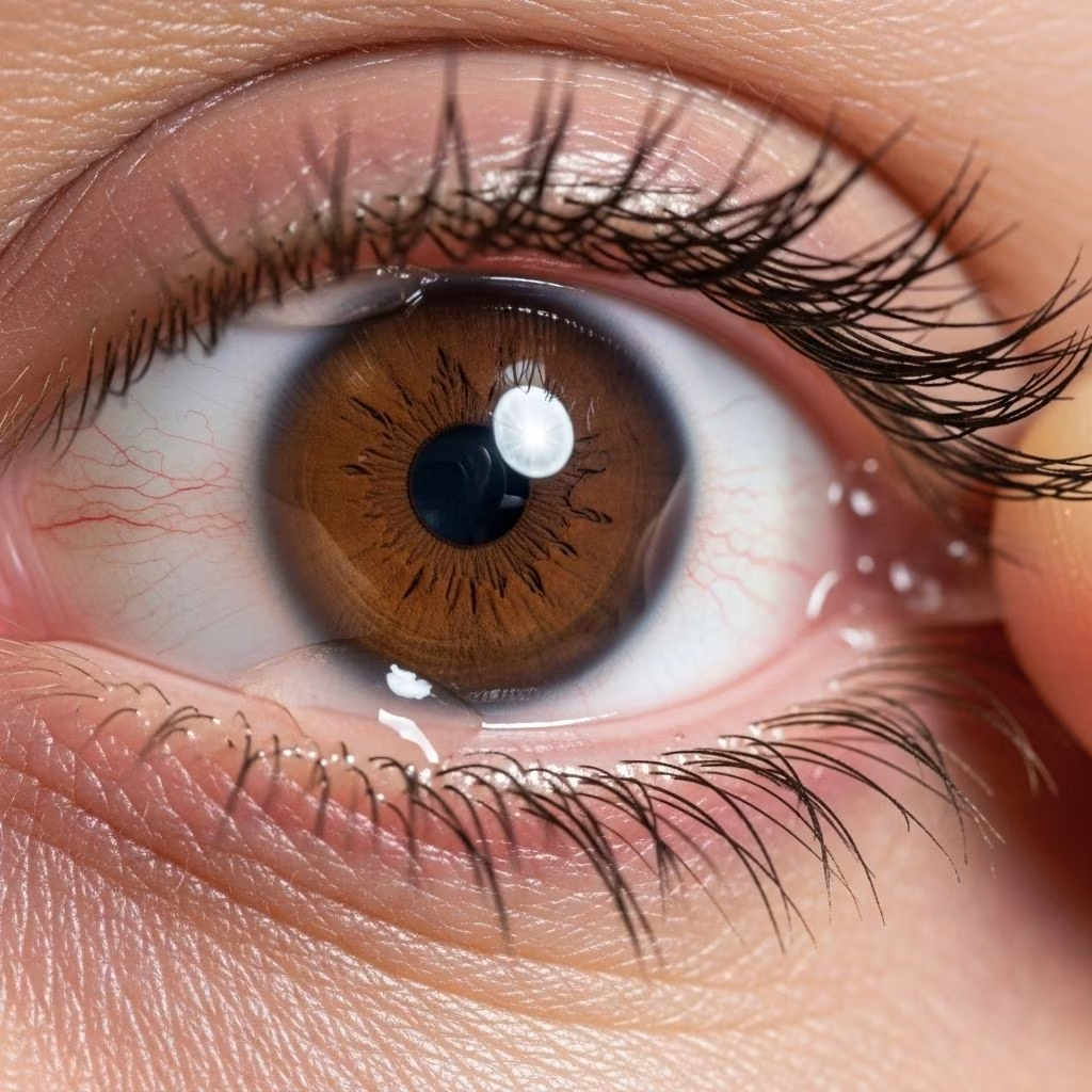
Conjunctivochalasis—sometimes abbreviated as CCh—is an eye condition where the conjunctiva, the thin, clear membrane that covers the white part of the eye and lines the inside of the eyelids, becomes loose and develops extra folds. These folds typically appear as wrinkled tissue accumulating over the lower edge of the eyeball, just above the lower eyelid.
- The word is derived from Greek: chalasis means “to slacken.”
- Most commonly affects adults over age 50 but may occur earlier due to eye inflammation, mechanical stress, or post-surgical changes.
- Frequently overlooked or misdiagnosed as dry eye disease due to overlapping symptoms.
What Does the Conjunctiva Do?
The conjunctiva creates a protective layer for the eye, hosts immune cells, helps keep the cornea moist and clear, and forms part of the tear film’s structure. Healthy conjunctiva contributes significantly to eye comfort and vision quality.
Symptoms of Conjunctivochalasis
Not all cases of conjunctivochalasis cause symptoms—in many people, extra conjunctival folds are found incidentally during routine eye examinations. However, when symptoms occur, they typically include:
- Dryness—a persistent dry sensation that does not improve with standard dry eye therapies.
- Irritation or burning—caused by redundant conjunctival tissue interfering with tear distribution.
- Foreign body sensation—feeling like there is something in the eye.
- Blurred or fluctuating vision—resulting from unstable tear film.
- Excessive tearing (epiphora)—because the tissue may block the drainage of tears rather than improve eye lubrication.
- Sensitivity to wind or light.
- Worsening discomfort when reading or blinking.
What Causes Conjunctivochalasis?
The causes of conjunctivochalasis are multiple and often interrelated. While aging is the most common underlying factor, several other mechanisms have been identified:
- Age-related degeneration—as we age, the connective tissue (elastic fibers) in the conjunctiva breaks down, causing it to lose firmness and sag.
- Chronic inflammation—conditions like blepharitis (inflammation of the eyelids), meibomian gland dysfunction, or persistent eye allergies can contribute to conjunctival breakdown.
- Mechanical stress—frequent rubbing of eyes or improper contact lens use may increase the risk.
- Previous eye surgery—procedures such as cataract or glaucoma surgery may lead to local tissue changes and loosening.
- Genetic factors and connective tissue disorders—Ehlers-Danlos Syndrome and similar diseases make conjunctival tissue more prone to looseness and wrinkling.
- Ultraviolet light exposure—chronic sunlight exposure may accelerate conjunctival aging.
Other Associated Conditions
- Blepharitis—Chronic eyelid inflammation
- Meibomian gland dysfunction—Disruption in lipid layer of tears
- Aqueous tear deficiency—Lack of watery layer in tears
How Does Conjunctivochalasis Affect the Eyes?
The redundant conjunctival folds can produce several distinct ocular disturbances:
- Disrupts tear lake—The normal “reservoir” of tears along the lower eyelid is interrupted, making the spread of tears uneven during blinking.
- Obstructs tear drainage—The extra tissue may block the tear ducts, causing watery eyes despite a sensation of dryness.
- Chronic irritation and inflammation—Due to physical friction between the folds and the lid margin, which may provoke more inflammation and perpetuate the problem.
Risk Factors for Developing Conjunctivochalasis
- Advancing age
- History of eye inflammation
- Chronic dry eye disease
- Previous ocular surgeries or trauma
- Repeated mechanical irritation
- Connective tissue disorders
Diagnosis of Conjunctivochalasis
A thorough eye examination by an ophthalmologist or optometrist is required to diagnose conjunctivochalasis. The process typically involves:
- History and symptoms—Identifying fluctuations in vision, unexplained dryness, or tearing that doesn’t respond to typical dry eye therapies.
- Slit-lamp examination—A high-magnification view reveals redundant folds or wrinkling of the conjunctiva, usually just above the lower eyelid margin.
- Tear breakup time (TBUT) and Schirmer’s test—May help distinguish between pure dry eye disease and conjunctivochalasis-related tear disruption.
Important: Many of the symptoms overlap with dry eye syndrome, making misdiagnosis common. Visualizing the presence of lid-parallel conjunctival folds is essential to distinguish conjunctivochalasis from other disorders.
How is Conjunctivochalasis Treated?
Treatment options depend on the severity of symptoms and the impact on quality of life:
1. Medical (Non-Surgical) Management
- Observation only—If asymptomatic or very mild, no treatment beyond monitoring is needed.
- Lubricating eye drops—Initial treatment involves using more viscous (gel-like) artificial tears, as these stay suspended over the irregular surface better than thin drops.
- Topical corticosteroids—Short-term topical steroid drops may be prescribed to reduce conjunctival inflammation. Long-term use is usually not recommended due to the risk of side effects.
- Artificial tear ointments—For night time lubrication, particularly if conjunctiva is exposed even when the eyes are closed.
- Eye patching at night—Can help prevent exposure and irritation during sleep in severe cases.
| Severity | Treatment Approach |
|---|---|
| Mild | Observe; lubricating drops as needed |
| Moderate | Lubricating gel drops; consider topical steroids |
| Severe | Ointment and eye patch at night; if ineffective, consider surgery |
2. Surgical Management
In cases where symptoms persist despite medical treatment or if excessive conjunctival folds significantly impair vision or comfort, surgical intervention may be necessary.
- Conjunctival resection—Excess conjunctiva is carefully excised to restore a smooth contour to the eye surface and to improve tear distribution.
- Amniotic membrane grafting—After removal, an amniotic membrane graft may be placed over the surgical area. This approach accelerates healing, reduces inflammation, and prevents scarring.
- Ocular surface reconstruction—More advanced procedures may include reconstructing the ocular surface or combining approaches for optimal tear film restoration.
Surgical treatment is typically safe and effective, often resulting in improved eye lubrication and relief from chronic irritation.
3. Prognosis After Treatment
- Most patients experience significant relief of symptoms following surgical correction.
- Recovery is generally swift, particularly with modern sutureless graft techniques.
- Long-term outcomes are excellent if contributing inflammation and lid disease are managed.
How to Prevent Conjunctivochalasis?
While not all cases are preventable, you can reduce your risk by:
- Avoiding unnecessary mechanical trauma to the eyes (e.g., rubbing or aggressive contact lens handling).
- Wearing sunglasses to limit ultraviolet exposure.
- Treating chronic eye inflammation or lid disease early.
- Following up regularly with your eye care provider, especially if you have existing ocular surface disorders or have undergone eye surgery.
Frequently Asked Questions (FAQs)
What’s the difference between conjunctivochalasis and dry eye disease?
Both conditions can cause eye dryness and irritation, but conjunctivochalasis specifically involves loose or wrinkled conjunctival tissue disrupting tear flow, whereas dry eye disease is more often due to insufficient or low-quality tear production. Sometimes, both occur together, making diagnosis and management more complex.
Can conjunctivochalasis be mistaken for other eye conditions?
Yes, it is often misdiagnosed as dry eye syndrome or allergy due to similar symptoms. The distinguishing feature is the visible redundant conjunctival folds upon slit-lamp exam.
Will lubricating eye drops cure conjunctivochalasis?
No, lubricating drops may offer symptom relief, especially in mild cases, but they do not correct the underlying tissue changes. Persistent or severe cases may need surgery.
Is the surgery for conjunctivochalasis painful?
Most surgical procedures are quick and performed under local anesthesia or light sedation. Discomfort is generally mild after the surgery and managed with drops or ointments.
How soon can I return to normal activities after surgery?
Recovery times vary, but many patients can resume daily activities within a week. Your eye doctor will give you specific guidelines based on the surgical technique used and the healing process.
Is conjunctivochalasis dangerous?
While it is rarely sight-threatening by itself, untreated severe conjunctivochalasis can lead to chronic inflammation, tear dysfunction, and in rare cases, susceptibility to infection or epithelial breakdown.
When to See an Eye Doctor
- If you experience persistent dry eye symptoms, blurry vision, or cannot tolerate your usual eye drops.
- If you develop a new foreign body sensation or tearing that does not improve with treatment.
- If you have a history of eye surgery and notice changes in your eye comfort or vision.
Early evaluation ensures accurate diagnosis and proper management to prevent unnecessary discomfort and maintain healthy vision.
Key Takeaways
- Conjunctivochalasis is a common, often overlooked cause of chronic eye discomfort in older adults.
- Symptoms overlap with dry eye but involve redundant conjunctival tissue visible on examination.
- Mild cases may not need treatment, but persistent or severe symptoms can be improved with medication or surgery.
- Modern surgical techniques offer safe, effective, and durable symptom relief.
- Regular eye exams are important for at-risk individuals, especially after age 50 or post eye surgery.
References
- https://www.theeyeassociates.com/services/dry-eye-disease/conjunctivochalasis-cch/
- https://www.thenewyorkeyedoctor.com/conjunctivochalasis-cch-.html
- https://www.ncbi.nlm.nih.gov/books/NBK576410/
- https://www.reviewofophthalmology.com/article/how-to-manage-conjunctivochalasis
- https://eyewiki.org/Conjunctivochalasis
- https://www.medicalnewstoday.com/articles/conjunctivochalasis
- https://www.eyeworld.org/2021/recognizing-understanding-and-treating-conjunctivochalasis/
- https://watsondryeyecenter.com/service/conjunctivochalasis/