

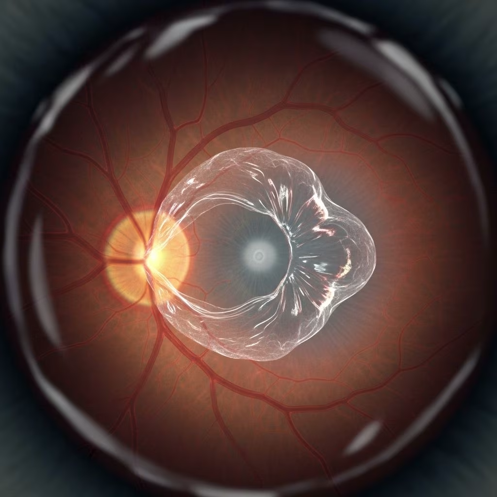
An epiretinal membrane (ERM), sometimes known as macular pucker or cellophane maculopathy, is a thin, transparent layer of fibrous tissue that can form on the macula—the central area of the retina responsible for detailed vision. While often mild or asymptomatic, ERMs can lead to visual distortion and blurriness for some people.
What Is an Epiretinal Membrane?
An epiretinal membrane is a semi-transparent, fibrocellular film or ‘scar tissue’ that develops on the internal surface of the retina, most often over the macula. The macula is crucial for tasks like reading, driving, and recognizing faces because it allows us to see fine details clearly.
This condition goes by several names, including:
- Macular pucker
- Cellophane maculopathy
- Premacular fibrosis
- Epi-macular membrane
Most ERMs result in mild or no symptoms and are identified during routine eye exams. In some cases, however, they can distort vision and interfere with daily activities.
How Common Is Epiretinal Membrane?
Epiretinal membranes are relatively common, especially as people age. They affect up to 8% of individuals over 60, with prevalence increasing with age.
While both eyes can develop ERM, it usually occurs in one eye. Having an ERM in one eye does not significantly raise the risk for developing it in the other eye.
Symptoms of Epiretinal Membrane
Many people with ERMs experience no symptoms. These membranes are often discovered incidentally during a comprehensive eye exam or through imaging such as optical coherence tomography (OCT). If symptoms do develop, they commonly include:
- Distorted vision (metamorphopsia): straight lines may appear wavy, crooked, or bent
- Blurry central vision
- Difficulty with tasks requiring fine vision (e.g., reading, recognizing faces)
- Objects appearing larger or smaller than normal (macropsia or micropsia)
- Double vision
- Light sensitivity (less common)
Peripheral (side) vision is not affected, and ERM does not cause complete blindness.
Causes and Risk Factors
An epiretinal membrane can occur without any apparent cause or be triggered by various eye conditions. The most frequent causes and risk factors are:
- Aging: Most ERMs are linked to age-related changes, specifically to posterior vitreous detachment (PVD), where the gel-like vitreous body detaches from the retina and releases cells that can form a membrane.
- Retinal conditions:
- Diabetic retinopathy
- Branch or central retinal vein occlusion
- Retinal tears or detachment
- Inflammatory diseases (e.g., uveitis)
- After retinal surgery or trauma
- PVD (Posterior Vitreous Detachment): Natural aging causes the vitreous to shrink and sometimes pull away from the retina, which can lead to cellular proliferation on the retinal surface.
- Ocular injury or previous eye surgery: Eye trauma or surgeries such as cataract operations can increase risk.
Though sometimes described as ‘scar tissue,’ ERM is not related to macular degeneration and rarely affects individuals under age 50.
How Does Epiretinal Membrane Affect Vision?
The impact of an ERM on vision varies widely. In early stages, vision may not be affected at all. In more developed cases, the membrane can shrink or contract and tug on the macula, causing the underlying retina to wrinkle, ‘pucker,’ or become uneven.
- Mild cases: No symptoms or barely noticeable visual distortion
- Moderate to severe cases: Blurred, wavy, or distorted central vision (metamorphopsia)
- Rarely: Decreased ability to see details, making reading and recognizing faces difficult
Distinguishing ERM from Other Macular Disorders
| Condition | Description | Main Features |
|---|---|---|
| Epiretinal Membrane (Macular Pucker) | Thin, fibrous layer on the macula | Visual distortion, blurring; doesn’t cause total blindness |
| Macular Degeneration | Degeneration of macula’s photoreceptors | Gradual central vision loss; can be severe |
| Macular Hole | Actual hole or break in the macula | Central vision loss, usually more severe than ERM |
Diagnosis of Epiretinal Membrane
Diagnosing ERM typically starts with a comprehensive eye exam by an ophthalmologist or optometrist. Key diagnostic methods include:
- Ophthalmoscopy: Direct visualization of the retina with a specialized microscope
- Optical Coherence Tomography (OCT): A noninvasive imaging test providing cross-sectional images of the retina to reveal wrinkling or thickening
- Retinal photography: High-resolution images of the retina surface
- Fluorescein angiography: Visualizes blood flow in retinal vessels, sometimes used to rule out other causes of visual symptoms
Not all individuals with ERM need extensive diagnostics, especially if the visual impact is minimal and not progressing.
How Is Epiretinal Membrane Treated?
Treatment decisions for ERM depend on the severity of the symptoms and their impact on daily life. Many cases require no intervention, only regular monitoring. If the membrane significantly distorts vision, surgery may be considered.
Observation and Monitoring
- People with mild or no symptoms often have their condition monitored through periodic eye exams.
- In most cases, ERMs do not worsen over time.
- Typically, the condition does not cause pain and does not lead to total loss of sight.
Surgical Treatment: Vitrectomy and Membrane Peeling
If significant blurring or distortion occurs, pars plana vitrectomy with membrane peeling is the standard treatment. This surgical procedure involves:
- Removing the vitreous gel from the eye (vitrectomy)
- Peeling the epiretinal membrane from the retinal surface using delicate microscopic forceps
Most people experience improvement in vision after surgery, though some may still have mild visual distortion. As with any surgery, there are risks to consider:
- Cataract development: Post-surgery cataracts can occur, especially in patients over 50
- Retinal detachment: Rare but more serious complication
- Infection or bleeding: Extremely rare ocular surgery risks
- Residual visual distortion: Some scar tissue or wrinkling may remain
Non-surgical treatments, like eye drops or medication, are ineffective for ERM.
Living with Epiretinal Membrane
Most people with ERM continue to perform everyday activities, including reading and driving, especially in mild or early cases. Practical advice for living well with ERM includes:
- Regular eye exams to monitor for progression
- Use of adequate lighting and magnifiers for reading
- Monitoring for symptoms such as increased blurriness or distortion
It’s important to report any sudden changes in vision to your eye care provider promptly.
When to See a Doctor
Consult an ophthalmologist or optometrist if you notice:
- Sudden or worsening visual distortions
- Difficulty performing daily visual tasks
- New symptoms, especially if they develop in only one eye
Early detection can help manage progression and maintain the highest possible quality of vision.
Frequently Asked Questions (FAQs)
What is the difference between an epiretinal membrane and macular degeneration?
Epiretinal membrane is a thin scar tissue that forms on the surface of the retina, causing distortion or blurring. Macular degeneration is a degeneration of the retinal tissue itself, often leading to gradual but potentially severe central vision loss.
Can an epiretinal membrane go away on its own?
Very rarely, an ERM may resolve by itself, usually if linked to an acute inflammatory or post-surgical event. Most membranes either remain stable or progress slowly, requiring only monitoring unless vision becomes affected.
Will I lose my vision completely if I have an epiretinal membrane?
No. ERM does not cause total blindness and does not affect peripheral (side) vision.
Is surgery always necessary for epiretinal membrane?
No. Most cases do not require surgery. Surgery is reserved for those experiencing significant vision loss or distortion affecting daily life.
Is epiretinal membrane related to macular holes?
Macular holes and ERM are distinct conditions, but the development of one can sometimes increase the risk of the other. Both affect the central retinal area, but ERM involves scar tissue on the surface, while a macular hole is a full-thickness defect.
Summary
Epiretinal membrane (macular pucker) is a common eye condition, especially among older adults. Most cases are mild and require only observation. When ERM causes marked visual disturbances, surgery can safely improve symptoms for most individuals. Regular eye exams and prompt reporting of new symptoms are essential for preserving vision and eye health.
For more information, always consult your eye doctor or a retina specialist.
References
- https://www.asrs.org/patients/retinal-diseases/19/epiretinal-membranes
- https://www.rnib.org.uk/your-eyes/eye-conditions-az/epiretinal-membrane/
- https://tanner-eyes.co.uk/epiretinal-membrane-causes-symptoms-and-treatment/
- https://www.msdmanuals.com/home/eye-disorders/retinal-disorders/epiretinal-membrane
- https://www.allaboutvision.com/conditions/epiretinal-membrane/
- https://www.webmd.com/eye-health/what-is-epiretinal-membrane
- https://www.mdfoundation.com.au/about-macular-disease/other-macular-conditions/epiretinal-membrane-macular-pucker/
- https://eyewiki.org/Epiretinal_Membrane
- https://www.mayoclinic.org/diseases-conditions/retinal-diseases/multimedia/img-20135596