

Retinal Holes: Causes, Symptoms, Treatment, and Prevention
The retina is a vital structure in your eye, converting light into electrical signals for the brain to interpret as vision. Retinal holes, especially macular holes, can disrupt this process and cause a significant loss of central vision. Understanding the causes, symptoms, treatment options, and preventive measures against retinal holes is crucial for maintaining lifelong eye health. This article comprehensively explores these topics and more, following the latest clinical insights and research.
What Is a Retinal Hole?
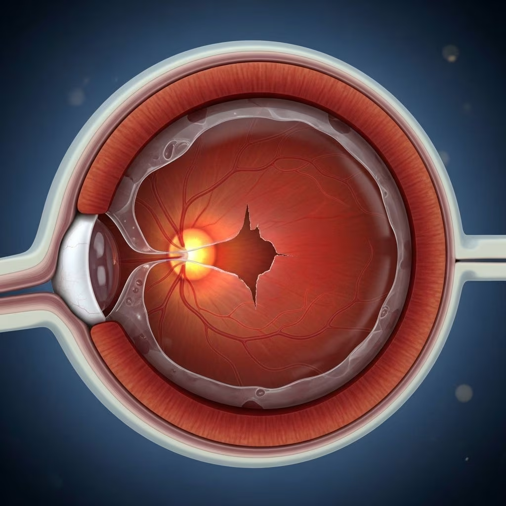
A retinal hole is a break or defect that occurs in the thin layer of tissue lining the back of your eye, known as the retina. One of the most well-known types is the macular hole, which occurs at the macula—the small, central part of the retina responsible for detailed central vision critical for reading, driving, and recognizing faces.
The retina transmits light signals to your brain which enables sight. If a hole develops, especially in the macula, it often leads to blur, dark spots, or distortion in the central vision field. While retinal holes are not exceedingly common, they can have a profound impact on quality of life if not promptly diagnosed and treated.
Anatomy: Retina, Macula, and How Vision Works
- Retina: Light-sensitive layer at the back of the eye covering roughly 65% of the eye’s interior surface.
- Macula: Small spot about the diameter of a pencil eraser located at the center of the retina. It provides sharp, central vision.
- Vitreous Humor: Gel-like substance filling the eyeball that can shrink or change over time, possibly tugging on the retina.
Healthy macular function is essential for detailed activities such as:
- Reading small print
- Driving
- Recognizing people’s faces
- Performing tasks requiring fine detail
Symptoms of Retinal (Macular) Hole
Symptoms usually develop gradually and primarily affect central vision in one eye, although both can be affected. Common symptoms include:
- Blurring of central vision
- Distortion, with straight lines appearing wavy
- Dark spot or area of missing vision in the center of the visual field
- Trouble with activities like reading or driving
- Difficulty recognizing faces
- Objects appearing smaller or more distant
Peripheral (side) vision typically remains unaffected unless accompanied by other retinal disease.
Severity and Progression
The degree to which vision is affected will depend on both the size of the hole and its exact location in the macula. The condition tends to worsen without intervention, leading to greater central vision loss over time.
Causes of Retinal and Macular Holes
Retinal holes—especially macular holes—mostly develop due to changes related to aging, but various factors and conditions may contribute:
- Aging (most common): The natural aging process causes the vitreous gel to shrink and tug at the retina, leading to a break in some cases.
- Vitreous traction: When the jelly-like vitreous humor pulls away from the retina abnormally, it can cause a tear or hole in the macula.
- Eye injury or trauma: A blow to the eye can cause a retinal hole, even in younger individuals.
- Diabetic eye disease: Diabetes-related changes can make the retina more vulnerable to holes.
- High myopia (severe nearsightedness): Increases the risk of retinal thinning and holes.
- Macular pucker: Formation of a scar tissue layer over the macula that can contract and warp, sometimes resulting in a hole.
- Rare factors: Retinal detachment or inflammation may also contribute in some cases.
Most cases are idiopathic (no obvious cause), especially in adults over 55.
Risk Factors
Certain populations are more prone to retinal or macular holes. Main risk factors include:
- Age: Most cases occur after age 55, especially after 60.
- Female gender: Women are affected more frequently than men.
- History of macular hole in the other eye: If you’ve had one, risk rises to 5-15% for the other.
- High myopia: Severe nearsightedness stretches and thins the retina.
- Previous eye injury: Trauma raises risk, especially at younger ages.
Diagnosis of a Retinal or Macular Hole
The diagnosis is made by a comprehensive eye exam where your eye doctor will use various tools to look at the retina. Diagnostic steps include:
- Dilated eye exam: Drops widen the pupil so the retina and macula can be closely examined.
- Slit-lamp examination: Allows close, magnified inspection of the retina’s structure to identify defects.
- Optical Coherence Tomography (OCT): A painless imaging test using light waves to produce high-resolution, cross-sectional images of the retina and macula. OCT is crucial for confirming the presence, size, and stage of a macular hole.
Macular Hole Staging
Macular holes are classified by stages, which guide treatment decisions:
| Stage | Description | Typical Vision Impact |
|---|---|---|
| Stage 1 | Impending or hole formation (foveal detachment) | Mild blurring/distortion |
| Stage 2 | Small full-thickness hole | Moderate distortion, central spot |
| Stage 3 | Large full-thickness hole | Significant central vision loss |
| Stage 4 | Hole with complete vitreous detachment | Severe central vision loss |
Treatment Options for Retinal (Macular) Holes
Treatment depends on the size, stage, and symptoms of the hole:
Observation (Wait-and-see Approach)
- Small, early-stage holes (especially Stage 1) may close spontaneously in a minority of cases (about 4% to 11.5%).
- If no significant vision loss, your doctor may choose to monitor the hole.
- Regular follow-up exams and OCT imaging are essential to monitor progression.
Medicated Eye Drops
- Research suggests that medicated drops—such as those targeting inflammation—may help close small macular holes without surgery for select patients, typically over several weeks.
Vitrectomy Surgery
- The gold standard for most full-thickness holes (Stages 2-4).
- What it involves: Removal of the vitreous gel (vitrectomy) and placement of a gas bubble inside the eye as a temporary support. This bubble helps the edges of the hole come together to heal.
- What to expect: Surgery is performed under local or general anesthesia, and most patients go home the same day.
- Postoperative Recovery: You may need to maintain a face-down (prone) position for several days to ensure the gas bubble is in optimal contact with the macular hole.
- Vision improvement takes place gradually over weeks to months after surgery.
- Surgical success rates are high, approaching 90% for suitable cases.
Other Treatments (Less Common)
- Ocriplasmin injection: An enzyme injection to dissolve proteins causing vitreous traction, rarely used but may be considered for early cases.
- Laser therapy is generally not used for macular holes.
Prognosis: What to Expect
With timely and appropriate care, many people with macular or retinal holes experience meaningful improvement or stabilization of vision. Key points:
- Small holes or early treatment have better visual outcomes.
- Delay in treatment or large holes lead to less favorable results.
- Peripheral vision is generally preserved even before and after treatment.
- There is a 5–15% risk of developing a macular hole in the other eye if you have one in one eye already.
Complications
- Persistent vision loss: Particularly if significant damage occurred before treatment.
- Retinal detachment: Uncommon but serious; requires emergency treatment.
- Recurrence of the hole after surgery (rare).
- Cataract development after vitrectomy is a common side effect that may require separate surgical treatment.
Prevention and Risk Reduction
While retinal holes often arise without clear warning or preventable cause, you can lower your risk or catch issues early by:
- Attending regular eye exams, especially after age 55 or if you have risk factors.
- Reporting any sudden vision changes to your healthcare provider immediately.
- Avoiding trauma by using protective eyewear during hazardous activities.
- Optimally managing diabetes and other systemic health conditions.
- Monitoring vision at home using an Amsler grid for early detection of wavy, missing, or distorted vision.
Living with a Retinal or Macular Hole
Adjustment to vision changes can be a challenge. Strategies include:
- Use magnifiers or electronic devices to aid reading and daily tasks.
- Improve lighting in living and work spaces.
- Rely more on the unaffected eye if vision loss is unilateral.
- Access low vision rehabilitation services for assistance.
Frequently Asked Questions (FAQs)
Q: What is the difference between a macular hole and a retinal tear?
A: A macular hole is a defect in the central part of the retina (macula), leading to loss of central vision. A retinal tear can happen anywhere along the retina, increasing risk for retinal detachment which affects peripheral vision.
Q: Is a macular hole the same as macular degeneration?
A: No. Macular holes are physical breaks in the retina, while macular degeneration is a chronic, progressive deterioration of the macula leading to vision loss. Their treatments and risk factors can differ significantly.
Q: Can a macular hole heal by itself?
A: A small percentage of early-stage holes may heal spontaneously, but most require surgical treatment for best visual outcomes.
Q: Will I lose all my vision if I have a macular hole?
A: Macular holes generally affect only central vision; most people retain their peripheral (side) vision.
Q: How soon should I seek treatment if I notice symptoms?
A: Prompt evaluation by an eye care professional is essential. Early intervention leads to better visual restoration and reduces the risk of complications.
Key Takeaways
- Retinal and macular holes significantly impact central vision but are treatable, especially with timely intervention.
- Pay attention to new vision changes and seek prompt eye evaluation.
- Modern treatments, including surgery and medicated drops for select cases, can restore or stabilize vision for most patients.
Maintaining regular eye exams and a proactive approach to eye health are the best defenses against the complications of retinal holes and other vision-impairing conditions.
References
- https://www.healthline.com/health/eye-health/macular-hole
- https://www.asrs.org/patients/retinal-diseases/4/macular-hole
- https://my.clevelandclinic.org/health/diseases/14208-macular-hole
- https://www.uchicagomedicine.org/forefront/research-and-discoveries-articles/2021/january/medication-closes-small-macular-holes
- https://eyewiki.org/Macular_Hole
- https://www.ncbi.nlm.nih.gov/books/NBK559200/
- https://jamanetwork.com/journals/jamaophthalmology/fullarticle/417557
- https://www.mayoclinic.org/medical-professionals/ophthalmology/news/macular-hole-repair-does-not-require-face-down-positioning/mac-20430610